. 2024 Nov 23;10(12):757.
doi: 10.3390/gels10120757.

Electrostatic Gelatin Nanoparticles for Biotherapeutic Delivery

Affiliations

Electrostatic Gelatin Nanoparticles for Biotherapeutic Delivery

Connor Tobo et al. Gels. .

Abstract

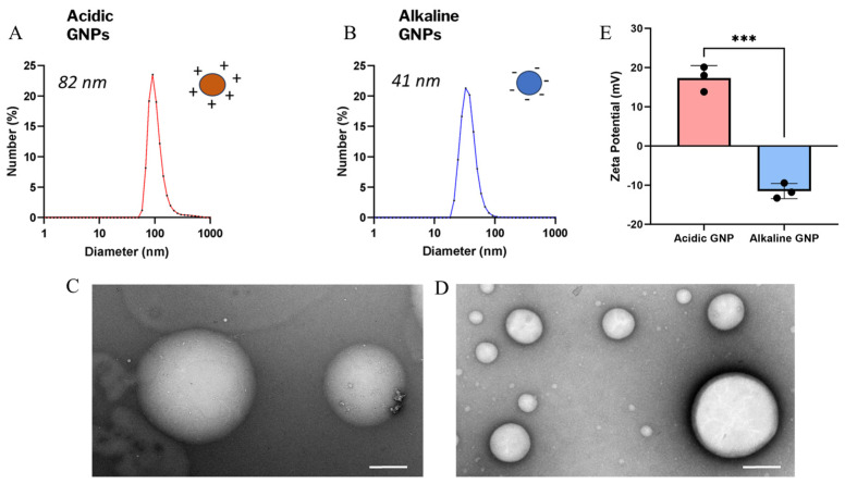
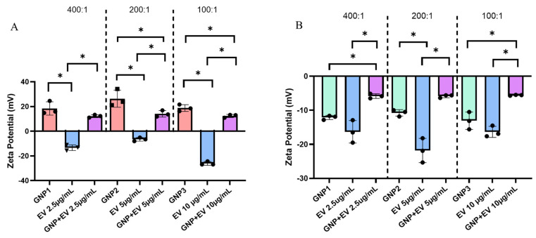
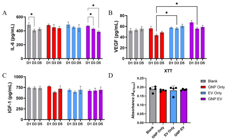
Biological agents such as extracellular vesicles (EVs) and growth factors, when administered in vivo, often face rapid clearance, limiting their therapeutic potential. To address this challenge and enhance their efficacy, we propose the electrostatic conjugation and sequestration of these agents into gelatin-based biomaterials. In this study, gelatin nanoparticles (GNPs) were synthesized via the nanoprecipitation method, with adjustments to the pH of the gelatin solution (4.0 or 10.0) to introduce either a positive or negative charge to the nanoparticles. The GNPs were characterized using dynamic light scattering (DLS), X-ray diffraction (XRD), Fourier-transform infrared spectroscopy (FTIR), and Transmission electron microscopy (TEM) imaging. Both positively and negatively charged GNPs were confirmed to be endotoxin-free and non-cytotoxic. Mesenchymal stem cell (MSC)-derived EVs exhibited characteristic surface markers and a notable negative charge. Zeta potential measurements validated the electrostatic conjugation of MSC-EVs with positively charged GNPs. Utilizing a transwell culture system, we evaluated the impact of EV-GNP conjugates encapsulated within a gelatin hydrogel on macrophage secretory activity. The results demonstrated the bioactivity of EV-GNP conjugates and their synergistic effect on macrophage secretome over five days of culture. In summary, these findings demonstrate the efficacy of electrostatically coupled biotherapeutics with biomaterials for tissue regeneration applications.

Keywords: electrostatic; extracellular vesicles; gelatin; nanoparticles.


Conflict of interest statement

The authors declare no conflicts of interest.

Figures

Figure 1
Figure 1
GNP size, morphology, and zeta potential characterization. The size distribution of acidic (A) and alkaline (B) GNPs is shown. TEM images show particle morphology and integrity of acidic (C) and alkaline (D) GNPs (scale bar = 200 nm). (E) Comparison of zeta potential of acidic GNPs vs. alkaline GNPs is presented. Data reported as mean ± standard deviation. A t-test demonstrates statistical significance (*** denotes p < 0.001, n = 3).
Figure 2
Figure 2
Powder XRD spectra comparing stock type-A gelatin (A) and acidic GNPs (B). FTIR spectra of both (C,E) type A and B gelatin and (D,F) type A and type B GNPs.
Figure 3
Figure 3
LDH Assay assessing cytotoxicity. No statistically significant differences were detected between GNP treatment groups and untreated media control (# denotes significance between all other groups, p < 0.001 (n = 3)).
Figure 4
Figure 4
MSC-EV size and surface marker characterization. (A) NTA data shows an average EV particle size range of 50–300 nm (n = 5). NTA for 1X PBS is also shown as negative control (n = 3). (B) An exo-check array reveals the presence of EV-specific markers, confirming the successful isolation of EVs. A marker for cellular contamination (GM130) was negative.
Figure 5
Figure 5
(A) Zeta potential measurements comparing acidic GNPs and EVs versus GNP+:EV conjugates. From left to right, GNP+:EV ratios tested include 400:1, 200:1, and 100:1. Each GNP and conjugate measurement maintained a consistent GNP concentration of 1.0 mg/mL while EV concentration was changed between groups. Different EV concentrations are specified (x-axis). (B) The zeta potential of alkaline GNPs, EVs, and their respectively combined solutions in the same ratios of 400:1, 200:1, and 100:1. Statistical comparisons between measurements of different GNP+ to EV ratios are not shown for ease of viewing. Data is represented as mean ± standard deviation (n = 3, statistical significance is denoted by * for p < 0.05 between groups for One-way ANOVA).
Figure 6
Figure 6
Release of GNP+:EV Conjugates and Their Temporal Effects on Bioactivity. (A) IL-6 secretion is significantly reduced in groups treated with GNP+:EV conjugates over the course of both three and five days, and it is also reduced on day three for cells cultured with control gels (B) Macrophages treated with EVs and GNP+:EV conjugates secreted significantly more VEGF than cells treated with gels containing only GNPs. (C) IGF-1 concentration is not significantly different amongst any groups. (D) No differences in cellular proliferation were noted between groups (n = 4, * denotes p < 0.05, a repeated measures two-way ANOVA was used for analysis).
Figure 7
Figure 7
GNP+:EV conjugation increases particle size and offers protection for improved retention of biological cargo.
Figure 8
Figure 8
GNP synthesis using the nanoprecipitation method and carbodiimide crosslinking prior to resuspension.

References

    1. Gang E.J., Jeong J.A., Hong S.H., Hwang S.H., Kim S.W., Yang I.H., Ahn C., Han H., Kim H. Skeletal myogenic differentiation of mesenchymal stem cells isolated from human umbilical cord blood. Stem Cells. 2004;22:617–624. doi: 10.1634/stemcells.22-4-617. - DOI - PubMed
    1. Seo B.F., Jung S.N. The Immunomodulatory Effects of Mesenchymal Stem Cells in Prevention or Treatment of Excessive Scars. Stem Cells Int. 2016;2016:6937976. doi: 10.1155/2016/6937976. - DOI - PMC - PubMed
    1. Caplan A.I. New MSC: MSCs as pericytes are Sentinels and gatekeepers. J. Orthop. Res. 2017;35:1151–1159. doi: 10.1002/jor.23560. - DOI - PubMed
    1. Guillamat-Prats R. The Role of MSC in Wound Healing, Scarring and Regeneration. Cells. 2021;10:1729. doi: 10.3390/cells10071729. - DOI - PMC - PubMed
    1. Lukomska B., Stanaszek L., Zuba-Surma E., Legosz P., Sarzynska S., Drela K. Challenges and Controversies in Human Mesenchymal Stem Cell Therapy. Stem Cells Int. 2019;2019:9628536. doi: 10.1155/2019/9628536. - DOI - PMC - PubMed

LinkOut - more resources