

"Chasing Rainbows" Beyond Kaposi Sarcoma's Dermoscopy: A Mini-Review
- PMID: 39727617
- PMCID: PMC11674653
- DOI: 10.3390/dermatopathology11040035
"Chasing Rainbows" Beyond Kaposi Sarcoma's Dermoscopy: A Mini-Review
Abstract
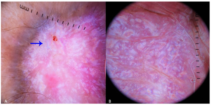
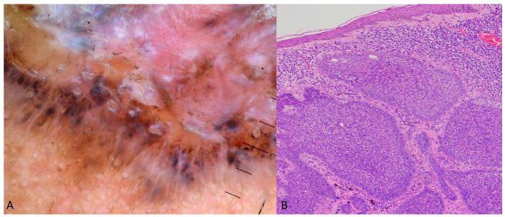
The dermoscopic rainbow pattern (RP), also known as polychromatic pattern, is characterized by a multicolored appearance, resulting from the dispersion of polarized light as it penetrates various tissue components. Its separation into different wavelengths occurs according to the physics principles of scattering, absorption, and interference of light, creating the optical effect of RP. Even though the RP is regarded as a highly specific dermoscopic indicator of Kaposi's sarcoma, in the medical literature, it has also been documented as an atypical dermoscopic finding of other non-Kaposi skin entities. We aim to present two distinct cases-a pigmented basal cell carcinoma (pBCC) and an aneurysmatic dermatofibroma-that exhibited RP in dermoscopy and to conduct a thorough review of skin conditions that display RP, revealing any predisposing factors that could increase the likelihood of its occurrence in certain lesions. We identified 33 case reports and large-scale studies with diverse entities characterized by the presence of RP, including skin cancers (Merkel cell carcinoma, BCC, melanoma, etc.), adnexal tumors, special types of nevi (blue, deep penetrating), vascular lesions (acroangiodermatitis, strawberry angioma, angiokeratoma, aneurismatic dermatofibromas, etc.), granulation tissue, hypertrophic scars and fibrous lesions, skin infections (sporotrichosis and cutaneous leishmaniasis), and inflammatory dermatoses (lichen simplex and stasis dermatitis). According to our results, the majority of the lesions exhibiting the RP were located on the extremities. Identified precipitating factors included the nodular shape, lesion composition and vascularization, skin pigmentation, and lesions' depth and thickness. These parameters lead to increased scattering and interference of light, producing a spectrum of colors that resemble a rainbow.
Keywords: dermatopathology; dermoscopy; polychromatic pattern; rainbow pattern; skin cancer.
Conflict of interest statement
The authors declare no conflicts of interest.
Figures

Similar articles
-
Dermoscopic rainbow pattern: A strong clue to malignancy or just a light show?North Clin Istanb. 2020 Aug 31;7(5):494-498. doi: 10.14744/nci.2020.32656. eCollection 2020. North Clin Istanb. 2020. PMID: 33163886 Free PMC article.
-
The rainbow pattern in dermoscopy: A zoom on nonkaposi sarcoma skin diseases.Biomed J. 2018 Jun;41(3):209-210. doi: 10.1016/j.bj.2018.04.004. Epub 2018 Jul 11. Biomed J. 2018. PMID: 30080661 Free PMC article.
-
Rainbow pattern in Kaposi's sarcoma under polarized dermoscopy: a dermoscopic pathological study.Br J Dermatol. 2009 Apr;160(4):801-9. doi: 10.1111/j.1365-2133.2008.08940.x. Epub 2008 Dec 1. Br J Dermatol. 2009. PMID: 19067686
-
Clinical and Dermoscopic Patterns of Basal Cell Carcinoma and Its Mimickers in Skin of Color: A Practical Summary.Medicina (Kaunas). 2024 Aug 24;60(9):1386. doi: 10.3390/medicina60091386. Medicina (Kaunas). 2024. PMID: 39336428 Free PMC article. Review.
-
The Dermoscopic Rainbow Pattern - A Review of the Literature.Acta Dermatovenerol Croat. 2019 Jun;27(2):111-115. Acta Dermatovenerol Croat. 2019. PMID: 31351506 Review.
Cited by
-
Exploring Pediatric Dermatology in Skin of Color: Focus on Dermoscopy.Life (Basel). 2024 Dec 4;14(12):1604. doi: 10.3390/life14121604. Life (Basel). 2024. PMID: 39768312 Free PMC article. Review.
-
Prognostic Factors and Long-Term Survival in Kaposi's Sarcoma Patients: Results from a 28-Year Retrospective Cohort.Medicina (Kaunas). 2025 Apr 14;61(4):724. doi: 10.3390/medicina61040724. Medicina (Kaunas). 2025. PMID: 40283016 Free PMC article.
References
Publication types
LinkOut - more resources
Full Text Sources
