Review
. 2024 Dec 1;9(12):733.
doi: 10.3390/biomimetics9120733.

Three-Dimensional Bioprinting for Retinal Tissue Engineering

Affiliations
Review

Three-Dimensional Bioprinting for Retinal Tissue Engineering

Kevin Y Wu et al. Biomimetics (Basel). .

Abstract

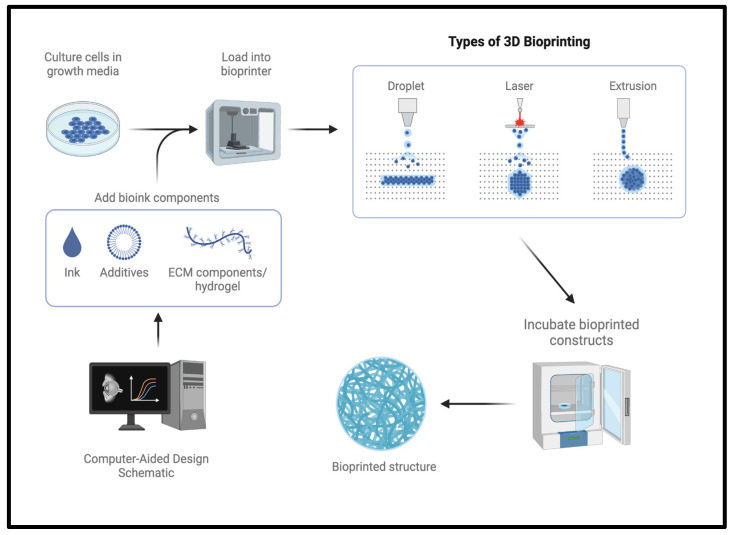
Three-dimensional bioprinting (3DP) is transforming the field of regenerative medicine by enabling the precise fabrication of complex tissues, including the retina, a highly specialized and anatomically complex tissue. This review provides an overview of 3DP's principles, its multi-step process, and various bioprinting techniques, such as extrusion-, droplet-, and laser-based methods. Within the scope of biomimicry and biomimetics, emphasis is placed on how 3DP potentially enables the recreation of the retina's natural cellular environment, structural complexity, and biomechanical properties. Focusing on retinal tissue engineering, we discuss the unique challenges posed by the retina's layered structure, vascularization needs, and the complex interplay between its numerous cell types. Emphasis is placed on recent advancements in bioink formulations, designed to emulate retinal characteristics and improve cell viability, printability, and mechanical stability. In-depth analyses of bioinks, scaffold materials, and emerging technologies, such as microfluidics and organ-on-a-chip, highlight the potential of bioprinted models to replicate retinal disease states, facilitating drug development and testing. While challenges remain in achieving clinical translation-particularly in immune compatibility and long-term integration-continued innovations in bioinks and scaffolding are paving the way toward functional retinal constructs. We conclude with insights into future research directions, aiming to refine 3DP for personalized therapies and transformative applications in vision restoration.

Keywords: 3D bioprinting; bioinks; biomimetics; biomimicry; microfluidics; organ-on-a-chip; regenerative medicine; retinal cells; retinal disease models; retinal tissue engineering; tissue scaffolds.


Conflict of interest statement

The authors declare no conflicts of interest.

Figures

Figure 1
Figure 1
Three-dimensional bioprinting process and types of bioprinting. Created in BioRender.
Figure 2
Figure 2
Keeling et al. [26] created reconstructed images of mice RPE 3D architecture (lateral view) showing apical microvilli (green) and nuclei (blue) with transparent cytoplasm allowing visualization of the convoluted basolateral Bruch’s membrane (yellow) with sub-RPE spaces (purple) and photoreceptors (light blue). Created in BioRender.
Figure 3
Figure 3
Deep to the outer pigmented aspect of the retina is the nine layers within the inner neural layer of the retina. The retina is located between the vitreous body and choroid [27]. Copyright certificate is CC by 3.0 license.
Figure 4
Figure 4
Retina structures cartoonized. Note: not all retinal layers are depicted in this figure. Created in BioRender.
Figure 5
Figure 5
Diagrammatic representation of the major requirements for a successful bioink. Created in BioRender.
Figure 6
Figure 6
Cartoonized rendering of the decellularization process for the development of decellularized ECM (dECM) biomaterial. The progressive loss of colour in this figure represents the loss of intracellular components in the decellularization process. The native retina tissue for which the ECM is derived is rendered in red, emblematic of the complex protein structures and intracellular environment supporting the native ECM. The final dECM product is rendered in gray, stripped of the native supportive proteins and growth-promoting intracellular environment. Created in BioRender.
Figure 7
Figure 7
Flow chart summarizing recent advancements of scaffold engineering in 3D retinal bioprinting. Many scaffolds are made with gellan gum (GG) as a base for its improved strength during the printing process. Created in Biorender.
Figure 8
Figure 8
Schematic representation of the oBRB. CC = choriocapillaris; TJ = tight junction. Created in Biorender.
Figure 9
Figure 9
Graphical representation of the drug-loaded combined bevacizumab/dexamethasone rod invention [94]. Created in Biorender.

Similar articles

Cited by

References

    1. Gu B.K., Choi D.J., Park S.J., Kim Y.-J., Kim C.-H. 3D Bioprinting Technologies for Tissue Engineering Applications. Adv. Exp. Med. Biol. 2018;1078:15–28. doi: 10.1007/978-981-13-0950-2_2. - DOI - PubMed
    1. Dell A.C., Wagner G., Own J., Geibel J.P. 3D Bioprinting Using Hydrogels: Cell Inks and Tissue Engineering Applications. Pharmaceutics. 2022;14:2596. doi: 10.3390/pharmaceutics14122596. - DOI - PMC - PubMed
    1. Ozbolat I.T. Scaffold-Based or Scaffold-Free Bioprinting: Competing or Complementing Approaches? J. Nanotechnol. Eng. Med. 2015;6:024701. doi: 10.1115/1.4030414. - DOI
    1. Ramadan Q., Zourob M. 3D Bioprinting at the Frontier of Regenerative Medicine, Pharmaceutical, and Food Industries. Front. Med. Technol. 2021;2:607648. doi: 10.3389/fmedt.2020.607648. - DOI - PMC - PubMed
    1. Mani M.P., Sadia M., Jaganathan S.K., Khudzari A.Z., Supriyanto E., Saidin S., Ramakrishna S., Ismail A.F., Faudzi A.A.M. A Review on 3D Printing in Tissue Engineering Applications. J. Polym. Eng. 2022;42:243–265. doi: 10.1515/polyeng-2021-0059. - DOI

LinkOut - more resources