Review
. 2024 Dec 18;9(12):771.
doi: 10.3390/biomimetics9120771.

Dehydrated Human Amnion-Chorion Membrane as a Bioactive Scaffold for Dental Pulp Tissue Regeneration

Affiliations
Review

Dehydrated Human Amnion-Chorion Membrane as a Bioactive Scaffold for Dental Pulp Tissue Regeneration

Sahng G Kim. Biomimetics (Basel). .

Abstract

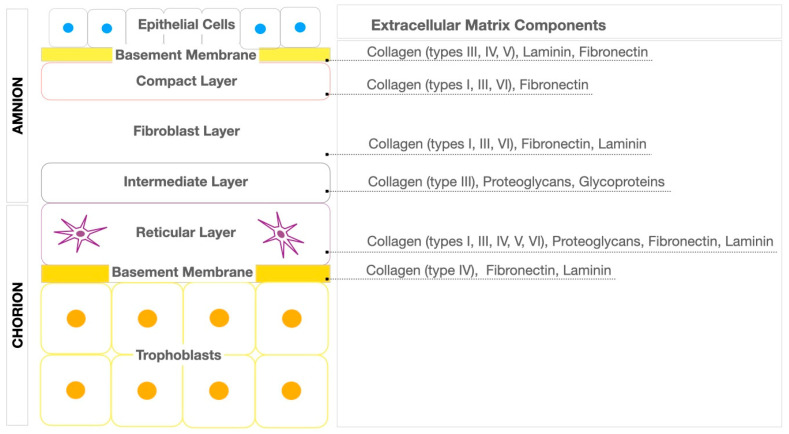
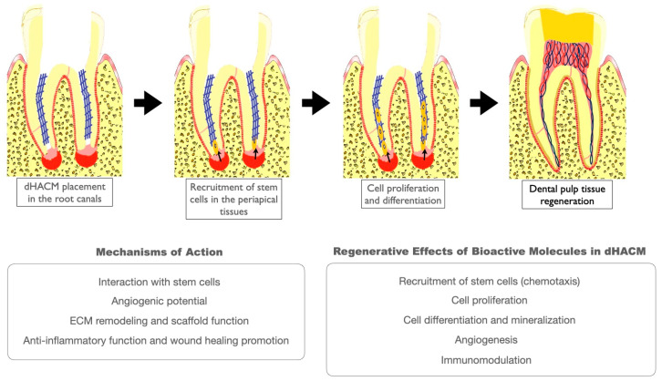
The dehydrated human amnion-chorion membranes (dHACMs) derived from the human placenta have emerged as a promising biomaterial for dental pulp regeneration owing to their unique biological and structural properties. The purpose of this review is to explore the potentials of dHACMs in dental pulp tissue engineering, focusing on their ability to promote cellular proliferation, differentiation, angiogenesis, and neurogenesis. dHACMs are rich in extracellular matrix proteins and growth factors such as TGF-β1, FGF2, and VEGF. They also exhibit significant anti-inflammatory and antimicrobial properties, creating an optimal environment for dental pulp regeneration. The applications of dHACMs in regenerative endodontic procedures are discussed, highlighting their ability to support the formation of dentin and well-vascularized pulp-like tissue. This review demonstrates that dHACMs hold significant potential for enhancing the success of pulp regeneration and offer a biologically based approach to preserve tooth vitality and improve tooth survival. Future research is expected to focus on conducting long-term clinical studies to establish their efficacy and safety.

Keywords: amnion–chorion membranes; bioactive scaffolds; biomaterials; dental pulp regeneration; regenerative endodontics.


Conflict of interest statement

The author declares no conflicts of interest.

Figures

Figure 1
Figure 1
Schematic diagram illustrating the structure of the amnion–chorion membrane and its detailed extracellular matrix components. The diagram highlights the distinct layers of the membrane—amnion and chorion—showing their unique composition and the types of collagen fibers, proteoglycans, glycoproteins, fibronectin, and laminin.
Figure 2
Figure 2
Cell homing-based pulp regeneration using dHACMs. dHACMs promote the recruitment of stem cells from the periapical tissue (e.g., stem cells of the apical papilla, bone marrow stem cells, periodontal ligament stem cells) and augment cellular events which are critical for pulp regeneration, such as angiogenesis, odontoblast differentiation, cell proliferation, and immunomodulation. Moreover, the membranes possess anti-inflammatory and antimicrobial properties and enhance wound healing and ECM remodeling through their function as a bioactive scaffold.
Figure 3
Figure 3
Clinical case using dHACMs for pulp regeneration. (A) Preoperative radiograph showing the maxillary right central incisor with a periapical lesion. The tooth was diagnosed with previously initiated therapy and symptomatic apical periodontitis (B) Postoperative radiograph after root canal disinfection, placement of dHACMs, and restoration. (C) Three-month follow-up showing a reduction in size of the periapical lesion. (D) Fourteen-month follow-up showing the complete resolution of the periapical lesion and the complete root formation.
Figure 4
Figure 4
Clinical case using dHACMs for vital pulp therapy. (A,D) Preoperative radiograph showing the mandibular left first molar with extensive caries involving the distal pulp horn. The tooth was diagnosed with symptomatic irreversible pulpitis and symptomatic apical periodontitis. (B,E) Postoperative radiograph after the removal of both caries and inflamed coronal pulp tissue, the placement of dHACMs, and restoration. (C,F) Three-month follow-up showing no periapical lesion. The tooth was negative to percussion and palpation and positive to electric pulp testing.

Similar articles

Cited by

References

    1. Kumar N., Maher N., Amin F., Ghabbani H., Zafar M.S., Rodríguez-Lozano F.J., Oñate-Sánchez R.E. Biomimetic Approaches in Clinical Endodontics. Biomimetics. 2022;7:229. doi: 10.3390/biomimetics7040229. - DOI - PMC - PubMed
    1. Sui B., Chen C., Kou X., Li B., Xuan K., Shi S., Jin Y. Pulp Stem Cell-Mediated Functional Pulp Regeneration. J. Dent. Res. 2019;98:27–35. doi: 10.1177/0022034518808754. - DOI - PubMed
    1. He L., Kim S.G., Gong Q., Zhong J., Wang S., Zhou X., Ye L., Ling J., Mao J.J. Regenerative Endodontics for Adult Patients. J. Endod. 2017;43:S57–S64. doi: 10.1016/j.joen.2017.06.012. - DOI - PubMed
    1. Kim S.G., Malek M., Sigurdsson A., Lin L.M., Kahler B. Regenerative endodontics: A comprehensive review. Int. Endod. J. 2018;51:1367–1388. doi: 10.1111/iej.12954. - DOI - PubMed
    1. Yang J., Yuan G., Chen Z. Pulp Regeneration: Current Approaches and Future Challenges. Front. Physiol. 2016;7:58. doi: 10.3389/fphys.2016.00058. - DOI - PMC - PubMed

LinkOut - more resources