Concave Magnetic-Responsive Hydrogel Discs for Enhanced Bioassays
- PMID: 39727861
- PMCID: PMC11674732
- DOI: 10.3390/bios14120596
Concave Magnetic-Responsive Hydrogel Discs for Enhanced Bioassays
Abstract
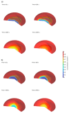
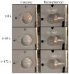
Receptor-based biosensors often suffer from slow analyte diffusion, leading to extended assay times. Moreover, existing methods to enhance diffusion can be complex and costly. In response to this challenge, we presented a rapid and cost-effective technique for fabricating concave magnetic-responsive hydrogel discs (CMDs) by straightforward pipetting directly onto microscope glass slides. This approach enables immediate preparation and customization of hydrogel properties such as porosity, magnetic responsiveness, and embedded particles and is adaptable for use with microarray printers. The concave design increased the surface area by 43% compared to conventional hemispherical hydrogels, enhancing diffusion rates and accelerating reactions. By incorporating superparamagnetic particles, the hydrogels become magnetically responsive, allowing for stirring within reagent droplets using magnets to improve mixing. Our experimental results showed that CMDs dissolved approximately 2.5 times faster than hemispherical ones. Numerical simulations demonstrated up to a 46% improvement in diffusion speed within the hydrogel. Particles with lower diffusion coefficients, like human antibodies, benefited most from the concave design, resulting in faster biosensor responses. The increased surface area and ease of fabrication make our CMDs efficient and adaptable for various biological and biomedical applications, particularly in point-of-care diagnostics where rapid and accurate biomarker detection is critical.
Keywords: bioassays; concave hydrogel discs; magnetic-responsive hydrogels.
Conflict of interest statement
The authors declare no conflict of interest.
Figures


Similar articles
-
Supramolecular Hydrogels for 3D Biosensors and Bioassays.Chemistry. 2024 Aug 19;30(46):e202400974. doi: 10.1002/chem.202400974. Epub 2024 Jul 30. Chemistry. 2024. PMID: 38871646 Review.
-
Hydrogel Nanoarchitectonics: An Evolving Paradigm for Ultrasensitive Biosensing.Small. 2022 Jul;18(26):e2107571. doi: 10.1002/smll.202107571. Epub 2022 May 27. Small. 2022. PMID: 35620959 Review.
-
Stimuli-bioresponsive hydrogels as new generation materials for implantable, wearable, and disposable biosensors for medical diagnostics: Principles, opportunities, and challenges.Adv Colloid Interface Sci. 2023 Jul;317:102920. doi: 10.1016/j.cis.2023.102920. Epub 2023 May 13. Adv Colloid Interface Sci. 2023. PMID: 37207377 Review.
-
DNA -based hydrogels for high-performance optical biosensing application.Talanta. 2022 Jul 1;244:123427. doi: 10.1016/j.talanta.2022.123427. Epub 2022 Mar 31. Talanta. 2022. PMID: 35390683
-
Injectable, porous, biohybrid hydrogels incorporating decellularized tissue components for soft tissue applications.Acta Biomater. 2018 Jun;73:112-126. doi: 10.1016/j.actbio.2018.04.003. Epub 2018 Apr 10. Acta Biomater. 2018. PMID: 29649634 Free PMC article.
References
-
- Kulshreshtha N.M., Shrivastava D., Bisen P.S. 14—Contaminant sensors: Nanotechnology-based contaminant sensors. In: Grumezescu A.M., editor. Nanobiosensors. Academic Press; Cambridge, MA, USA: 2017. pp. 573–628. - DOI
-
- Goodrum R., Aggarwal R.T., Li H. Gold-nanoparticle-embedded membrane (GEM) for highly sensitive multiplexed sandwich immunoassays. Sens. Actuators B Chem. 2024;410:135731. doi: 10.1016/j.snb.2024.135731. - DOI
-
- Wu C., Du L., Zou L., Zhao L., Huang L., Wang P. Recent advances in taste cell- and receptor-based biosensors. Sens. Actuators B Chem. 2014;201:75–85. doi: 10.1016/j.snb.2014.04.021. - DOI
-
- Nakano S., Konishi H., Morii T. Chapter Nine—Receptor-based fluorescent sensors constructed from ribonucleopeptide. In: Chenoweth D.M., editor. Methods in Enzymology. Volume 641. Academic Press; Cambridge, MA, USA: 2020. pp. 183–223. - PubMed
MeSH terms
Substances
Grants and funding
LinkOut - more resources
Full Text Sources

