Review
. 2024 Dec 6;14(12):597.
doi: 10.3390/bios14120597.

Nanolabels Prepared by the Entrapment or Self-Assembly of Signaling Molecules for Colorimetric and Fluorescent Immunoassays

Affiliations
Review

Nanolabels Prepared by the Entrapment or Self-Assembly of Signaling Molecules for Colorimetric and Fluorescent Immunoassays

Ning Xia et al. Biosensors (Basel). .

Abstract

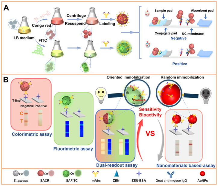
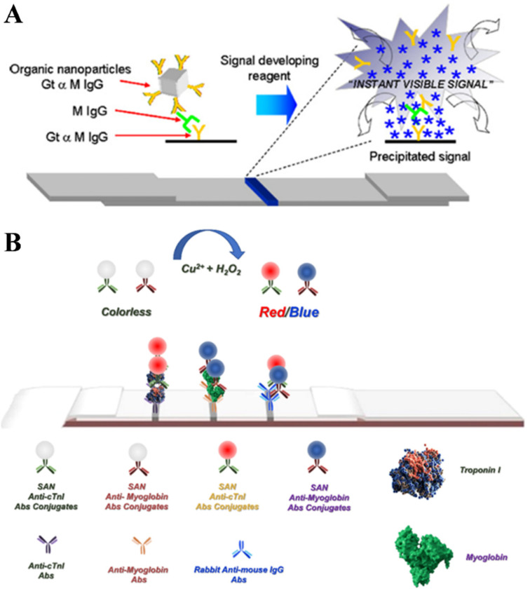
Nanomaterials have attracted significant attention as signal reporters for immunoassays. They can directly generate detectable signals or release a large number of signaling elements for readout. Among various nanolabels, nanomaterials composed of multiple signaling molecules have shown great potential in immunoassays. Generally, signaling molecules can be entrapped in nanocontainers or self-assemble into nanostructures for signal amplification. In this review, we summarize the advances of signaling molecules-entrapped or assembled nanomaterials for colorimetric and fluorescence immunoassays. The nanocontainers cover liposomes, polymers, mesoporous silica, metal-organic frameworks (MOFs), various nanosheets, nanoflowers or nanocages, etc. Signaling molecules mainly refer to visible and/or fluorescent organic dyes. The design and application of immunoassays are emphasized from the perspective of nanocontainers, analytes, and analytical performances. In addition, the future challenges and research trends for the preparation of signaling molecules-entrapped or assembled nanolabels are briefly discussed.

Keywords: immunoassays; nanocontainers; nanomaterials; self-assembly; signaling molecules.


Conflict of interest statement

The authors declare no conflicts of interest.

Figures

Figure 4
Figure 4
(A) (a) Preparation of MOFs NH2-MIL-53(Al) and (b) schematic illustration of competitive FIA of AFB1 [81]. Copyright 2019 American Chemical Society. (B) Schematic illustration of the synthetic procedure of MILL-88@TcP nanozyme-based detection probe (top) and the procedure of this developed N-ELISA for S. typhimurium detection in milk (bottom) [89]. Copyright 2024 Elsevier.
Figure 5
Figure 5
(A) Schematic representation of MPDA@TP-linked immunosorbent assay (MLISA) for α-fetoprotein (AFP) on anti-AFP capture antibody (CAb)-modified microplate using anti-AFP detection antibody (DAb)-labeled MPDA@TP with a sandwich-type immunoreaction mode [92]. Copyright 2018 American Chemical Society. (B) The synthesis of UiO, UiOL, UiOL@AIEgens, and UiOL@AIEgens-mAbs probe (a), and the UiOL@AIEgens-based POC LFIS for visual and quantitative dual-modal detection of AFB1 (b) [96]. Copyright 2024 Elsevier.
Figure 9
Figure 9
(A) Workflow of porphyrin nanoparticle-based signal amplification sandwich assays for the detection of biomolecules [112]. Copyright 2016 American Chemical Society. (B) Scheme of sandwich-type TLISA for the detection of IL-6 [115]. Copyright 2019 American Chemical Society.
Figure 1
Figure 1
(A) Schematic of the liposome-amplified plasmonic immunoassay (LAPIA). The detection steps include the capture (a) and recognition (b) of the target, attachment of streptavidin (c), coupling of biotin-conjugated cysteine-contained liposomes (d), breakdown of the liposomes (e), and release of cysteine to trigger the aggregation of AuNPs (f) [27]. Copyright 2015 American Chemical Society. (B) Schematic illustration of signal-on competitive-type colorimetric immunoassay for the detection of streptomycin (STR) on monoclonal anti-STR antibody-coated microplate using glucose-loaded liposome as the signal tracer labeled with STR-bovine serum albumin (BSA) conjugate: (a) competitive-type immunoreaction and (b) glucose oxidase (GOx)-triggered the change of the Fe(II)-Phen system in the absorbance and visual color by the reaction of the produced H2O2 with iron(II) [41]. Copyright 2018 Elsevier.
Figure 2
Figure 2
Schematic presentation of the heterogeneous sandwich immunoassay with PSP for loading load C153, hemin, or microperoxidase MP11 based on different signal generation strategies and photo/chemiluminescence detection [51]. Copyright 2024 American Chemical Society.
Figure 3
Figure 3
(A) (a) Synthesis and derivatization of TP@PEI/Ab2-MSNs and (b) steps of the enzyme-free immunosorbent assay of PSA using for amplified colorimetric detection in a 96-well plate [63]. Copyright 2018 American Chemical Society. (B) Schematic illustration of the magnetic bead (MB)-based colorimetric immunoassay of PSA by the redox cycling with Ab2-MSN-PQQ as the nanolabel [64]. Copyright 2019 Elsevier. (C) Schematic illustration of the fluorescence immunoassay based on target-induced competitive displacement reaction between glucose and mannose for Con A accompanying cargo (rhodamine B) release from magnetic mesoporous silica nanoparticles (MMSNs) [65]. Copyright 2013 American Chemical Society.
Figure 6
Figure 6
(A) (a) The preparation process for the signal label of PP-Ab2-cC3N4, and (b) schematic illustration of the PILISA for the detection of CEA in 96-well PS plates [100]. Copyright 2017 Elsevier. (B) Schematic of AuNF@Fluorescein@SA preparation, and AuNF@Fluorescein@SA-based dual-mode fluorescent and colorimetric immunoassay [101]. Copyright 2018 Elsevier.
Figure 7
Figure 7
(A) Synthetic procedure for SAD carriers and their applications in ICAs. (B) Comparison of SAD-ICAs with three modes and a traditional nanomaterial—ICA (take AuNPs as an example)—for the detection of ZEN [102]. Copyright 2021 American Chemical Society.
Figure 8
Figure 8
(A) Principle of immunoassay using antigen-decorated perylene microparticles [109]. Copyright 2000 Elsevier. (B) Principle of a sandwich fluorescent immunoassay using nanocrystalline fluorescein diacetate (FDA) conjugates [103]. Copyright 2004 American Chemical Society.
Figure 10
Figure 10
(A) Schematic diagram illustrating the principle of using organic nanoparticles as biolabels for immunodipsticks [119]. Copyright 2011 Elsevier. (B) Schematic representation of the strategy of integrating an SAN-LFA for the detection of cardiac biomarkers [120]. Copyright 2016 American Chemical Society.

Similar articles

Cited by

References

    1. Farka Z., Jurik T., Kovar D., Trnkova L., Skladal P. Nanoparticle-based immunochemical biosensors and assays: Recent advances and challenges. Chem. Rev. 2017;117:9973–10042. doi: 10.1021/acs.chemrev.7b00037. - DOI - PubMed
    1. Pan R., Li G., Liu S., Zhang X., Liu J., Su Z., Wu Y. Emerging nanolabels-based immunoassays: Principle and applications in food safety. TrAC Trend. Anal. Chem. 2021;145:116462. doi: 10.1016/j.trac.2021.116462. - DOI
    1. Farka Z., Brandmeier J.C., Mickert M.J., Pastucha M., Lacina K., Skladal P., Soukka T., Gorris H.H. Nanoparticle-based bioaffinity assays: From the research laboratory to the market. Adv. Mater. 2024;36:e23076532024. doi: 10.1002/adma.202307653. - DOI - PubMed
    1. Kokkinos C., Economou A. Emerging trends in biosensing using stripping voltammetric detection of metal-containing nanolabels—A review. Anal. Chim. Acta. 2017;961:12–32. doi: 10.1016/j.aca.2017.01.016. - DOI - PubMed
    1. Svechkarev D., Mohs A.M. Organic fluorescent dye-based nanomaterials: Advances in the rational design for imaging and sensing applications. Curr. Med. Chem. 2019;26:4042–4064. doi: 10.2174/0929867325666180226111716. - DOI - PMC - PubMed

LinkOut - more resources