. 2024 Nov 22;22(12):526.
doi: 10.3390/md22120526.

Discovery and Characterization of an Atypical Crustin Antimicrobial Peptide from Pollicipes pollicipes

Affiliations

Discovery and Characterization of an Atypical Crustin Antimicrobial Peptide from Pollicipes pollicipes

Wei Zhang et al. Mar Drugs. .

Abstract

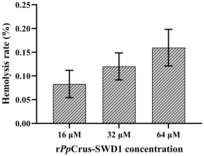
Crustins are a family of antimicrobial peptides (AMPs) that play a pivotal role in the innate immune system of crustaceans. The discovery of novel AMPs from natural sources is crucial for expanding our current database of these peptides. Here, we identified and characterized a novel member of the crustin family, named PpCrus-SWD1, derived from Pollicipes pollicipes. PpCrus-SWD1 consists of 138 amino acids and contains eight cysteine residues that form a conserved 'four-disulfide core' structure. Our recombinant PpCrus-SWD1 (rPpCrus-SWD1) exhibited potent inhibitory activity against three Gram-positive bacteria (Staphylococcus aureus, Bacillus sp. T2, and Streptococcus agalactiae) and six Gram-negative bacteria (Aeromonas hydrophila, Escherichia coli, Vibrio anguillarum, Vibrio alginolyticus, Vibrio parahemolyticus, and Acinetobacter sp. L3), with minimum inhibitory concentrations ranging from 16 to 64 μM. Furthermore, rPpCrus-SWD1 demonstrated binding affinity towards both bacteria and pathogen-associated molecular patterns (PAMPs), and damaged bacterial barrier. Additionally, it effectively inhibited alkaline protease activity in S. aureus and V. alginolyticus strains. These findings highlight the potential utility of this newly discovered crustin as an effective alternative to antibiotics.

Keywords: Pollicipes pollicipes; antibacterial mechanism; antibiotic; antimicrobial peptides; crustin.


Conflict of interest statement

The authors declare no conflicts of interest.

Figures

Figure 1
Figure 1
Schematic representation (not to scale) of the structural organization of the four crustin types (I to VII) found in crustaceans. CRD, cysteine-rich region; GRD, glycine-rich region; SRD, serine-rich region; LRD, leucine-rich region; WAP domain, whey acidic protein domain; SS, signal peptide.
Figure 2
Figure 2
Sequence alignments and phylogenetic and structural analysis of PpCrus-SWD1. (A) Alignment of PpCrus-SWD1 with type III crustins (shown in Supplementary Table S2). The consensus residues are shaded black; the residues that are 100% identical among the aligned sequences are shaded blue; the residues that are ≥75% identical among the aligned sequences are shaded pink, the residues that are ≥50% identical among the aligned sequences are shaded cyan (B) Phylogenetic analysis of PpCrus-SWD1 (red star) homologs. The phylogenetic tree was constructed with MEGA-X using the neighbor-joining method. Numbers beside the internal branches indicate bootstrap values based on 1000 replications. The crustins’ information used to construct the evolutionary tree is in Supplementary Tables S3 and S4. (C) The predicted structure of PpCrus-SWD1 was built using Alphafold2. The cysteines in the WAP domain are shown in Pink, and the β-pleated sheet is shown in blue.
Figure 3
Figure 3
The acquisition process and MS spectrum analysis of rPpCrus-SWD1. (A) SDS-PAGE analysis of recombinant PpCrus-SWD1 (rPpCrus-SWD1) expressed with a SUMO tag in E. coli. Lane M, protein marker; lane 1, total protein obtained from E. coli without induction; lane 2, total protein obtained from E. coli with IPTG induction. (B) His-SUMO-PpCrus-SWD1 purified with nickel column chromatography. Lane M, protein marker; lane 1, protein not caught by the nickel column; lane 2, equilibration buffer; lane 3, eluent with 20 mM Imidazole; lane 4, eluent with 50 mM Imidazole; lane 5, eluent with 100 mM Imidazole, lane 6, eluent with 200 mM Imidazole; lane 7, eluent with 500 mM Imidazole. (C) SDS-PAGE analysis of rPpCrus-SWD1 without SUMO tag. Lane M, protein marker; lane 1, His-SUMO-PpCrus-SWD1 after treatment with SUMO enzyme; lane 2, rPpCrus-SWD1 obtained after removing SUMO tag; the red arrow points to the band of rPpCrus-SWD1. (D) Alignment of mass spectrometry results with rPpCrus-SWD1 sequence. (E) MS spectrum of “ETCVGPGCGPLSSQLVAACENLPAPHPCTFFTCPPGKSCADR”. The red, blue, and black lines are the y ions, b ions, and noise signals detected by mass spectrometry, respectively.
Figure 4
Figure 4
Molecular dynamics simulation. (A) The radii of gyration (Rg) of PpCrus-SWD1 in aqueous solution and membrane. (B) The root–mean–square distances (RMSDs) of PpCrus-SWD1 in aqueous solution and membrane. (C) The root–mean–square fluctuations (RMSFs) of PpCrus-SWD1 in aqueous solution and membrane. (D) PpCrus-SWD1 and membrane binding simulation (initial stage). (E) PpCrus-SWD1 and membrane binding simulation (100 ns). (F) PpCrus-SWD1 and membrane binding simulation (200 ns).
Figure 5
Figure 5
Microorganism-binding activity and PAMP-binding activity of rPpCrus-SWD1. (A) Microorganism-binding activity. rPpCrus-SWD1 was detected by Western Blot assay after treatment with bacteria (Gram+ and Gram). rPpCrus-SWD1 was taken as a positive control. Up panel, elution fractions; bottom panel, final pellet fractions. (B) Lipoteichoic acid binding activity. (C) Peptidoglycan binding assay. (D) Lipopolysaccharide binding assay. rPpCrus-SWD1 was detected by ELISA. rPpCrus-SWD1 was taken as a positive control. SUMO tag was taken as a negative control. LTA, Lipoteichoic acid; PGN, Peptidoglycan; LPS, Lipopolysaccharide. *, Compared with control (0 μM), p-value < 0.01.
Figure 6
Figure 6
Morphological and cell membrane integrity changes of the bacterial cells treated with rPpCrus-SWD1. Morphological changes of the bacterial cells treated with rPpCrus-SWD1. About 106 CFU·mL−1 bacteria were incubated with 2 × MIC of rPpCrus-SWD1 for 2 h and observed under scan electron microscopy. PBS was used as a control. The scales are 2 μm. At the red arrow, the suspected bacterial content can be seen flowing out, and the cell surface shrinks.
Figure 7
Figure 7
The effect of rPpCrus-SWD1 on bacterial cell membrane integrity. About 1 × 10⁶ CFU·mL−1 bacteria were incubated with 2×MIC of rAaCrus1 for 2 h. PI: The cells were stained with PI and observed for PI uptake with a fluorescence microscope; BF: The bright field image. The scales are 50 μm.
Figure 8
Figure 8
Ability to inhibit protease activity and DNA binding activity of rPpCrus-SWD1. (A) Ability to inhibit the alkaline protease activity of S. aureus; (B) Ability to inhibit the alkaline protease activity of V. alginolyticus; (C) Ability to inhibit the neutral protease activity of S. aureus; (D) Ability to inhibit the neutral protease activity of V. alginolyticus; (E) Binding activity of rPpCrus-SWD1 to plasmid DNA; lane M, marker; lane 1, negative control (BSA); lane 2, 8 μM rPpCrus-SWD1 treatment; lane 3, 16 μM rPpCrus-SWD1 treatment; lane 4, 32 μM rPpCrus-SWD1 treatment; *, Compared with control (16 μM BSA), p-value < 0.01.
Figure 9
Figure 9
Hemolytic activity of rPpCrus-SWD1 on sheep red blood cells. The hemolysis rate of 1× PBS (pH 7.4) treatment was 0%, and that of 0.2% Triton X-100 treatment was 100%. There was no significant difference in hemolysis rate among the three concentration treatment groups (p-value > 0.05).

Similar articles

Cited by

References

    1. Yadav N., Chauhan V.S. Advancements in Peptide-Based Antimicrobials: A Possible Option for Emerging Drug-Resistant Infections. Adv. Colloid Interface Sci. 2024;333:103282. doi: 10.1016/j.cis.2024.103282. - DOI - PubMed
    1. Khan R.T., Rasool S., Sharma V., Khan S.S. Prevention and Potential Remedies for Antibiotic Resistance: Current Research and Future Prospects. Front. Microbiol. 2024;15:1455759. doi: 10.3389/fmicb.2024.1455759. - DOI - PMC - PubMed
    1. Chen P., Ye T., Li C., Praveen P., Hu Z., Li W., Shang C. Embracing the Era of Antimicrobial Peptides with Marine Organisms. Nat. Prod. Rep. 2024;41:331–346. doi: 10.1039/D3NP00031A. - DOI - PubMed
    1. Aisenbrey C., Marquette A., Bechinger B. Antimicrobial Peptides. Springer; Singapore: 2019. The Mechanisms of Action of Cationic Antimicrobial Peptides Refined by Novel Concepts from Biophysical Investigations; pp. 33–64. - PubMed
    1. Li W., Separovic F., O’Brien-Simpson N.M., Wade J.D. Chemically Modified and Conjugated Antimicrobial Peptides against Superbugs. Chem. Soc. Rev. 2021;50:4932–4973. doi: 10.1039/D0CS01026J. - DOI - PubMed

MeSH terms

LinkOut - more resources