. 2024 Nov 25;22(12):529.
doi: 10.3390/md22120529.

Therapeutic Potential of Oligo-Fucoidan in Mitigating Peritoneal Dialysis-Associated Fibrosis

Affiliations

Therapeutic Potential of Oligo-Fucoidan in Mitigating Peritoneal Dialysis-Associated Fibrosis

Yu-Wei Chen et al. Mar Drugs. .

Abstract

Peritoneal dialysis (PD) serves as a home-based kidney replacement therapy with increasing utilization across the globe. However, long-term use of high-glucose-based PD solution incites repeated peritoneal injury and inevitable peritoneal fibrosis, thus compromising treatment efficacy and resulting in ultrafiltration failure eventually. In the present study, we utilized human mesothelial MeT-5A cells for the in vitro experiments and a PD mouse model for in vivo validation to study the pathophysiological mechanisms underneath PD-associated peritoneal fibrosis. High-glucose PD solution (Dianeal 4.25%, Baxter) increased protein expression of mesothelial-mesenchymal transition (MMT) markers, such as N-cadherin and α-SMA in MeT-5A cells, whereas it decreased catalase expression and stimulated the production of reactive oxygen species (ROS). Furthermore, macrophage influx and increased serum pro-inflammatory cytokines, such as IL-1β, MCP-1, and TNF-α, were observed in the PD mouse model. Interestingly, we discovered that oligo-fucoidan, an oligosaccharide extract from brown seaweed, successfully prevented PD-associated peritoneal thickening and fibrosis through antioxidant effect, downregulation of MMT markers, and attenuation of peritoneal and systemic inflammation. Hence, oligo-fucoidan has the potential to be developed into a novel preventive strategy for PD-associated peritoneal fibrosis.

Keywords: MMT; fibrosis; mesothelial–mesenchymal transition; oligo-fucoidan; peritoneal dialysis.


Conflict of interest statement

The authors declare no conflicts of interest. The funders had no role in the design of the study; in the collection, analyses, or interpretation of data; in the writing of the manuscript; or in the decision to publish the results.

Figures

Figure 1
Figure 1
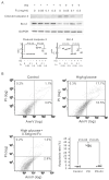
Influence of oligo-fucoidan (Fc) on the signal transduction of epithelial–mesenchymal transition and inflammation in MeT-5A cells. The cells were pretreated with Fc for 30 min and then cultured in high glucose (HG, containing 60 mM glucose) or normal medium. Protein expression was analyzed by Western blotting. α-SMA, E-cadherin, and N-cadherin are the markers of epithelial–mesenchymal transition. Phospho-JNK and TNF-α are the markers of inflammation. The relative increase in protein bands is also presented as a chart. Results are expressed as mean ± SD (n = 3).
Figure 2
Figure 2
The protective effect of oligo-fucoidan (Fc) on high-glucose-induced apoptosis in MeT-5A cells. The cells were pretreated with Fc for 30 min and then cultured in high glucose (HG, containing 60 mM glucose) or normal medium (control). (A) The Western blots of cleaved caspases-3 and Bcl-2. Cleaved caspases-3 and Bcl-2 are the markers of apoptosis. The relative increase in protein bands is also presented as a chart. Results are expressed as mean ± SD (n = 3). (B) The representative flow cytometric plots of cell apoptosis. In each plot, the lower left quadrant represents viable cells, the upper left quadrant necrotic cells, the lower right quadrant early apoptotic cells, and the upper right quadrant necrotic or late apoptotic cells. The apoptotic rate is also presented as a chart. Results are expressed as mean ± SD (n = 3). High-glucose-induced apoptosis in MeT-5A cells, which was inhibited by 0. 5 mg/mL Fc. AnnV: Annexin V-FITC, PI: propidium iodide.
Figure 3
Figure 3
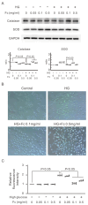
Effects of oligo-fucoidan (Fc) on high-glucose-induced reactive oxygen species (ROS) generation in MeT-5A cells. MeT-5A cells were pretreated with Fc for 30 min and then cultured in high glucose (HG, containing 60 mM glucose) or normal medium. (A) The Western blots of catalase and SOD. Catalase and SOD are the markers of antireactive oxygen species. The relative increase in protein bands is also presented as a chart. Results are expressed as mean ± SD (n = 3). (B) Representative images by fluorescent staining with 2′,7′-dichlorofluorescin (DCF). The image represents a combination of visible light microscopy and fluorescence microscopy images of MeT-5A cells. Green signifies the fluorescence of DCF when stained with ROS. (C) The intensity of DCF fluorescence in the cells measured by a fluorescence microplate reader. Fluorescence intensities of cells are shown as the relative intensity of experimental groups compared with untreated control cells. High-glucose-induced ROS in MeT-5A cells, which was inhibited by 0.1 and 0.5 mg/mL Fc. Data are shown in mean ± S.D. (n = 5).
Figure 4
Figure 4
Masson trichrome staining of the parietal peritoneum of PD mice. PD mice received 4.25% dextrose dialysate or without (control) and were then treated with oligo-fucoidan (Fc) orally (100 mg/kg/d). Average parietal peritoneum thickness in PD mice was presented as a chart. Dialysate increased parietal peritoneum thickness in PD mice, which was inhibited by Fc. Data are shown in mean ± S.D. (n = 5).
Figure 5
Figure 5
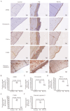
Morphology of the parietal peritoneum of PD mice with or without the oral oligo-fucoidan (Fc) treatment. Sections of paraffin-embedded mouse parietal peritoneum were stained by IHC using antibodies for α-SMA, fibronectin, TNF-α, F4/80, and cleaved caspase-3 (A). Red arrows highlight F4/80-positive macrophages and cleaved caspase-3-positive cells in the parietal peritoneum. Charts also display the positively stained areas in the peritoneum for α-SMA (B), fibronectin (C), and TNF-α (D), as well as the positively stained cells for F4/80 (E) and cleaved caspase-3 (F). Data are presented as the mean ± SD from 3 mice in each group.
Figure 6
Figure 6
The reduction effect of oligo-fucoidan (Fc) on serum TNF-α, MCP-1, and IL-1β in PD mice. PD mice received 4.25% glucose dialysate or without (control) and were then treated with Fc orally (100 mg/kg/d). Blood was collected from each mouse to measure serum TNF-α (A), MCP-1 (B), and IL-1β (C). Dialysate induced serum TNF-α, MCP-1, and IL-1β in PD mice, which was inhibited by Fc. The results are expressed as the mean ± SD (n = 3).

Similar articles

References

    1. GBD Chronic Kidney Disease Collaboration Global, regional, and national burden of chronic kidney disease, 1990–2017: A systematic analysis for the Global Burden of Disease Study 2017. Lancet. 2020;395:709–733. doi: 10.1016/S0140-6736(20)30045-3. - DOI - PMC - PubMed
    1. Li P.K.-T., Chow K.M., Van de Luijtgaarden M.W.M., Johnson D.W., Jager K.J., Mehrotra R., Naicker S., Pecoits-Filho R., Yu X.Q., Lameire N. Changes in the worldwide epidemiology of peritoneal dialysis. Nat. Rev. Nephrol. 2017;13:90–103. doi: 10.1038/nrneph.2016.181. - DOI - PubMed
    1. Thurlow J.S., Joshi M., Yan G., Norris K.C., Agodoa L.Y., Yuan C.M., Nee R. Global Epidemiology of End-Stage Kidney Disease and Disparities in Kidney Replacement Therapy. Am. J. Nephrol. 2021;52:98–107. doi: 10.1159/000514550. - DOI - PMC - PubMed
    1. Raoofi S., Pashazadeh Kan F., Rafiei S., Hoseinipalangi Z., Rezaei S., Ahmadi S., Masoumi M., Noorani Mejareh Z., Roohravan Benis M., Sharifi A., et al. Hemodialysis and peritoneal dialysis-health-related quality of life: Systematic review plus meta-analysis. BMJ Support. Palliat. Care. 2023;13:365–373. doi: 10.1136/bmjspcare-2021-003182. - DOI - PubMed
    1. Teitelbaum I. Peritoneal Dialysis. N. Engl. J. Med. 2021;385:1786–1795. doi: 10.1056/NEJMra2100152. - DOI - PubMed

MeSH terms

LinkOut - more resources