. 2024 Nov 21;15(12):355.
doi: 10.3390/jfb15120355.

Impact of Particle Size and Sintering Temperature on Calcium Phosphate Gyroid Structure Scaffolds for Bone Tissue Engineering

Affiliations

Impact of Particle Size and Sintering Temperature on Calcium Phosphate Gyroid Structure Scaffolds for Bone Tissue Engineering

Romina Haydeé Aspera-Werz et al. J Funct Biomater. .

Abstract

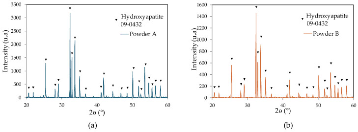
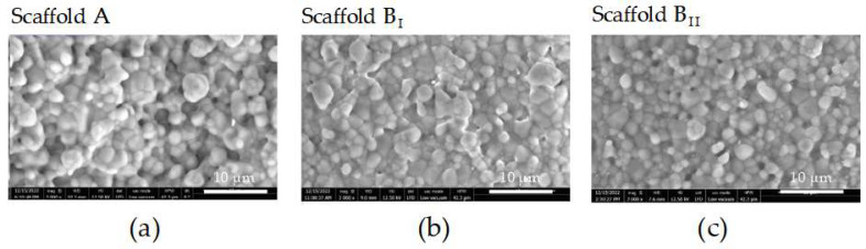
Due to the chemical composition and structure of the target tissue, autologous bone grafting remains the gold standard for orthopedic applications worldwide. However, ongoing advancements in alternative grafting materials show that 3D-printed synthetic biomaterials offer many advantages. For instance, they provide high availability, have low clinical limitations, and can be designed with a chemical composition and structure comparable to the target tissue. This study aimed to compare the influences of particle size and sintering temperature on the mechanical properties and biocompatibility of calcium phosphate (CaP) gyroid scaffolds. CaP gyroid scaffolds were fabricated by 3D printing using powders with the same chemical composition but different particle sizes and sintering temperatures. The physicochemical characterization of the scaffolds was performed using X-ray diffractometry, scanning electron microscopy, and microtomography analyses. The immortalized human mesenchymal stem cell line SCP-1 (osteoblast-like cells) and osteoclast-like cells (THP-1 cells) were seeded on the scaffolds as mono- or co-cultures. Bone cell attachment, number of live cells, and functionality were assessed at different time points over a period of 21 days. Improvements in mechanical properties were observed for scaffolds fabricated with narrow-particle-size-distribution powder. The physicochemical analysis showed that the microstructure varied with sintering temperature and that narrow particle size distribution resulted in smaller micropores and a smoother surface. Viable osteoblast- and osteoclast-like cells were observed for all scaffolds tested, but scaffolds produced with a smaller particle size distribution showed less attachment of osteoblast-like cells. Interestingly, low attachment of osteoclast-like cells was observed for all scaffolds regardless of surface roughness. Although bone cell adhesion was lower in scaffolds made with powder containing smaller particle sizes, the long-term function of osteoblast-like and osteoclast-like cells was superior in scaffolds with improved mechanical properties.

Keywords: bone graft; calcium phosphate scaffolds; osteoblast-like cells; osteoclast-like cells; sintering.


Conflict of interest statement

Authors Islam Bouakaz, Catherine Bronne, Elisabeth Cobraiville, and Gregory Nolens were employed by the company Cerhum. The remaining authors declare that the research was conducted in the absence of any commercial or financial relationships that could be construed as a potential conflict of interest.

Figures

Figure 1
Figure 1
Scaffold with cylindrical shape and gyroid structure. (a) Top view. (b) Side view.
Figure 2
Figure 2
Powder composition determined by X-ray diffractometry (XRD). Representative XRD curve for (a) powder A and (b) powder B.
Figure 3
Figure 3
Mechanical characterization of the three scaffolds tested. (a) Maximum force, (b) maximum stress, (c) displacements at maximum load, and (d) flexural strength were analyzed on scaffolds generated with powder A sintering at 1230 °C [Scaffold A] or powder B sintering at 1250 °C and 1210 °C [scaffolds BI and BII, respectively]. The data are presented as the mean, standard error of the mean, and all data points. Data were analyzed by the Kruskal–Wallis test followed by Dunn’s multiple comparisons. p-values are classified as * p < 0.05; *** p < 0.001; and **** p < 0.0001 for comparison between scaffold B and scaffold A and as # p < 0.05 for comparison within scaffold B.
Figure 4
Figure 4
Surface topographies of the three scaffolds analyzed by scanning electron microscopy (SEM). (a) Scaffold generated with powder A and sintering at 1230 °C. (b) Scaffold generated with powder B and sintering at 1250 °C. (c) Scaffold generated with powder B and sintering at 1210 °C (scale bar 10 µm).
Figure 5
Figure 5
SCP-1 cell attachment, number of live cells, and proliferation on three scaffolds tested. SCP-1 cells were seeded and cultured on scaffolds A, BI, and BII for 21 days. (a) Attached SCP-1 cells on scaffolds compared to cultured polystyrene. Number of live SCP-1 cells were analyzed after 24 h, 48 h, 7 days, 14 days, and 21 days by total DNA levels (b) and visualized by esterase activity (c) using calcein-AM (green) and nuclear staining using Hoechst 33342 (blue) (scale bar 2000 µm). Each measure was conducted at least three independent times in triplicate. The data are presented as the mean, standard error of the mean, and all data points. Data were analyzed by the Kruskal–Wallis test followed by Dunn’s multiple comparisons (a) or a two-way analysis of variance test followed by Tukey’s multiple comparisons (b). p-values are classified as ** p < 0.01; *** p < 0.001; and **** p < 0.0001 for comparison between scaffold B and scaffold A and as ## p < 0.01; ### p < 0.001; and #### p < 0.0001 for comparison within scaffold B.
Figure 6
Figure 6
SCP-1 osteogenic differentiation potential on three scaffolds tested. SCP-1 cells were seeded and cultured under osteogenic condition on scaffolds A, BI, and BII for 21 days. (a) Metabolic activity of SCP-1 cells were analyzed after 24 h, 48 h, 7 days, 14 days, and 21 days by mitochondrial activity as relative fluorescence units (RFU). (b) Alkaline phosphatase (AP) activity normalized to DNA of SCP-1 cells were analyzed after 7 days, 14 days, and 21 days as relative absorbance units (RAU). (c) Procollagen type I N-propeptide (PINP) supernatant levels were determined after 21-day osteogenic culture. Each measure was conducted at least three independent times in duplicate. The data are presented as the mean, standard error of the mean, and all data points. Data were analyzed by a two-way analysis of variance test followed by Tukey’s multiple comparisons. p-values are classified as * p < 0.05; ** p < 0.01; and **** p < 0.0001 for comparison between scaffold B and scaffold A and as # p < 0.05; ## p < 0.01; ### p < 0.001; and #### p < 0.0001 for comparison within scaffold B.
Figure 7
Figure 7
THP-1 cell attachment and number of live cells on three scaffolds tested. THP-1 cells were seeded and cultured on scaffolds A, BI, and BII for 24 h. (a) Attached SCP-1 cells on scaffolds compared to cultured polystyrene. Number of live THP-1 cells were visualized after 24 h by esterase activity (b) using calcein-AM (green) and nuclear staining using Hoechst 33342 (blue) (scale bar 2000 µm). Each measure was conducted at least three independent times in triplicate. The data are presented as the mean, standard error of the mean, and all data points. Data were analyzed by the Kruskal–Wallis test followed by Dunn’s multiple comparisons. p-values are classified as **** p < 0.0001 for comparison between scaffold B and scaffold A.
Figure 8
Figure 8
Bone cell viability and proliferation in co-cultures on the three scaffolds tested. THP-1 and SCP-1 were seeded and co-cultured on scaffolds A, BI, and BII for 21 days. Quantification of total DNA (a) and mitochondrial activity by resazurin conversion (b) in bone co-cultures after 7 days, 14 days, and 21 days. Number of live bone co-cultures were visualized by esterase activity (c) using calcein-AM (green) and nuclear staining with Hoechst 33342 (blue) (scale bar 2000 µm). Each measure was conducted at least three independent times in triplicate. The data are presented as the mean, standard error of the mean, and all data points. Data were analyzed by two-way analysis of variance test followed by Tukey’s multiple comparisons. p-values are classified as **** p < 0.0001 for comparison between scaffold B and scaffold A and as # p < 0.05 for comparison within scaffold B.
Figure 9
Figure 9
Osteoblast- and osteoclast-like cell function in co-cultures on the three scaffolds tested. THP-1 and SCP-1 were seeded and co-cultured on scaffolds A, BI, and BII for 21 days. (a) Alkaline phosphatase (AP), (b) carbonic anhydrase II (CAII), and (c) tartrate-resistant acid phosphatase (TRAP) activity normalized to DNA of bone co-cultures were analyzed after 7 days, 14 days, and 21 days as relative absorbance units (RAU). (d) Procollagen type I N-propeptide (PINP) and collagen type I N-telopeptide (NTX) supernatant levels were determined after a 21-day culture. Each measure was conducted at least three independent times in duplicate. The data are presented as the mean or standard error of the mean. Data were analyzed by a two-way analysis of variance test followed by Tukey’s multiple comparisons. p-values are classified as **** p < 0.0001, ** p < 0.01, and * p < 0.05 for comparison between scaffold B and scaffold A and as ## p < 0.01 for comparison within scaffold B.

References

    1. Florencio-Silva R., Sasso G.R., Sasso-Cerri E., Simões M.J., Cerri P.S. Biology of Bone Tissue: Structure, Function, and Factors That Influence Bone Cells. BioMed Res. Int. 2015;2015:421746. doi: 10.1155/2015/421746. - DOI - PMC - PubMed
    1. Šromová V., Sobola D., Kaspar P. A Brief Review of Bone Cell Function and Importance. Cells. 2023;12:2576. doi: 10.3390/cells12212576. - DOI - PMC - PubMed
    1. Datta H.K., Ng W.F., Walker J.A., Tuck S.P., Varanasi S.S. The cell biology of bone metabolism. J. Clin. Pathol. 2008;61:577–587. doi: 10.1136/jcp.2007.048868. - DOI - PubMed
    1. Huang S., Jin M., Su N., Chen L. New insights on the reparative cells in bone regeneration and repair. Biol. Rev. Camb. Philos. Soc. 2021;96:357–375. doi: 10.1111/brv.12659. - DOI - PubMed
    1. Tarantino U., Cariati I., Greggi C., Gasbarra E., Belluati A., Ciolli L., Maccauro G., Momoli A., Ripanti S., Falez F., et al. Skeletal System Biology and Smoke Damage: From Basic Science to Medical Clinic. Int. J. Mol. Sci. 2021;22:6629. doi: 10.3390/ijms22126629. - DOI - PMC - PubMed

LinkOut - more resources