. 2024 Dec 2;15(12):363.
doi: 10.3390/jfb15120363.

Photodynamic Therapy Using IR-783 Liposomes for Advanced Tongue and Breast Cancers in Humans

Affiliations

Photodynamic Therapy Using IR-783 Liposomes for Advanced Tongue and Breast Cancers in Humans

Yasuo Komura et al. J Funct Biomater. .

Abstract

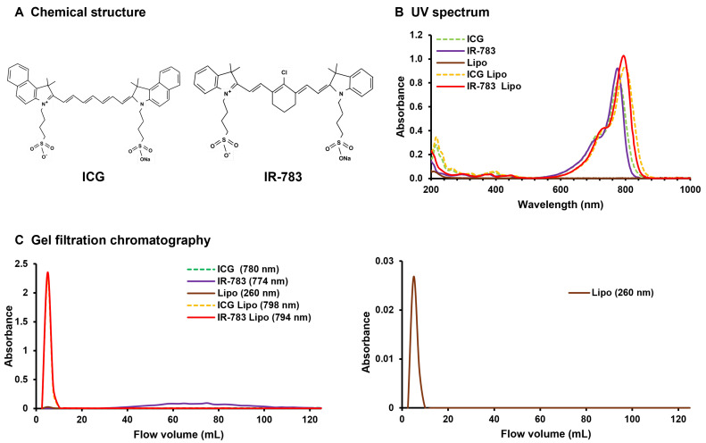
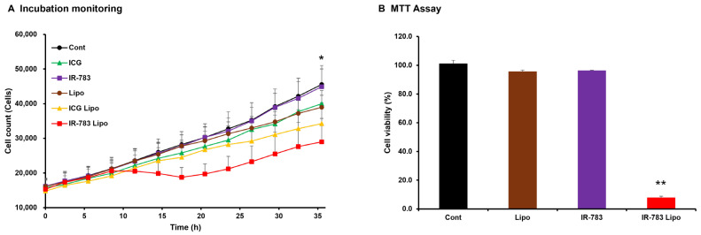
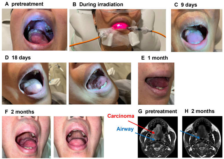
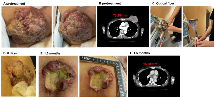
Photodynamic therapy (PDT) is a minimally invasive treatment that elicits tumor apoptosis using laser light exclusively applied to the tumor site. IR-783, a heptamethine cyanine (HMC) dye, impedes the proliferation of breast cancer cells, even without light. Although studies have investigated the efficacy of IR-783 in cell and animal studies, its efficacy in clinical settings remains unknown. Therefore, we aimed to determine the efficacy of PDT using IR-783 liposomes. An HMC dye, excited by long-wavelength infrared light and with high tissue permeability, was used for PDT after liposomization to enhance tumor tissue accumulation. PDT was performed using IR-783 in two patients with either tongue or breast cancer, one each. IR-783 liposomes inhibited cell proliferation in tongue cancer cells even when not excited by light. Tumor size was markedly reduced in both cases, with no significant adverse events. Furthermore, the patient with tongue cancer exhibited improved respiratory, swallowing, and speech functions, which were attributed not only to the shrinkage of the tumor but also to the improvement in airway narrowing. In conclusion, PDT using IR-783 liposomes effectively reduces tumor size in tongue and breast cancers.

Keywords: IR-783; breast cancer; heptamethine cyanine dyes; indocyanine green; liposome; lung cancer cells; photodynamic therapy; tongue cancer.


Conflict of interest statement

Shintarou Kimura, Yumi Hirasawa and Tomoko Katagiri are employees of StateArt Inc., Tokyo, Japan. The remaining authors declare that the research was conducted in the absence of any commercial or financial relationships that could be construed as a potential conflict of interest.

Figures

Figure 1
Figure 1
(A) Chemical structure of ICG and IR-783. (B) UV spectrum results. The light-grey dotted line depicts ICG in isolation, whereas the straight line represents IR-783. The dark grey straight line represents liposomes alone, the black dotted line represents ICG liposomes, and the straight line represents IR-783 liposomes. The absorbance peaks of both ICG and IR-783 tend to shift to the right when liposomes are used. (C) Gel filtration chromatography results. The light-grey dotted line depicts ICG in isolation, whereas the straight line represents IR-783. The dark-grey line represents liposomes alone, the black dotted line represents ICG liposomes, and the straight line represents IR-783 liposomes. The left side shows all liposome types, whereas the right side shows single liposomes with low absorbance values. ICG could not be quantified at the limit of detection, and IR-783 was only sporadically detected in the latter half of the sample owing to its small size. Both liposomized ICG and IR-783 are observed at the same locations as the standalone liposomes rather than at the sites where they are detected independently. This finding confirms that ICG and IR-783 are liposomized. ICG: indocyanine green; Lipo: liposomes.
Figure 2
Figure 2
(A) Result of real-time measurement of HCC827 cell proliferation over 36 h using an incubation monitoring system. HMC liposomes were treated with cells at equivalent concentrations to the respective HMC (10 μM), while controls and liposomes were treated at the same volume as the HMC (100 μL). The white circles represent the control group (5% glucose), while the black circles indicate the liposome group. The white triangles represent the ICG group, and the black triangles indicate the liposome group. The white squares represent the IR-783 group, and the black squares indicate the IR-783 liposome group. The vertical axis represents the number of cells, while the horizontal axis depicts time. Significant discrepancies in cellular proliferation after 36 h were determined exclusively between the control and IR-783 liposome groups. (B) Results of MTT assay for measuring the viability of HCC827 cells treated with IR-783 and IR-783 liposomes at 10 μM of IR-783 equivalent after 48 h. The controls and liposomes were treated with 100 μL, which was the same volume as that used for IR-783 and IR-783 liposomes. The cells treated with IR-783 liposomes show significantly lower viability compared with the control cells. The results were analyzed via one-way analysis of variance with Bonferroni’s post-test and are expressed as the mean ± standard deviation (n = 5, * p < 0.05, ** p < 0.01). ICG: indocyanine green; Lipo: liposomes.
Figure 3
Figure 3
(A) Images showing the patient with tongue cancer prior to photodynamic therapy (PDT) using IR-783. (B) The patient undergoing infrared light irradiation. A large tumor is visible on the right middle part of the tongue. (C) Image obtained 9 days following the initial intervention, showing a reduction in tumor size. (D) Image obtained 18 days following the initial intervention, showing that the tumor in the upper portion of the tongue was no longer discernible, and only an ulcerative lesion in the right side of the tongue was visible. (E,F) Images obtained 1 and 2 months following the initial intervention, respectively. No lesions were identified, and no observable tendency for recurrence was noted. (G) Magnetic resonance imaging (MRI) of the patient prior to PDT using IR-783. (H) MRI of the patient 2 months after the initial intervention. The red line denotes the tumor tissue, while the blue line represents the airway. After 2 months, the tumor was no longer discernible on the MRI, and the stenotic airway was recovered.
Figure 4
Figure 4
(A) Pretreatment image of the tumor in the breast cancer patient. A large-sized tumor was observed in the left breast. (B) Pretreatment CT image. (C) Image of the irradiation equipment used in PDT. Five optical fibers for irradiation were grouped together in a washer and wrapped with wrapping film. (D) Image captured 7 days after the first treatment. The tumor was slightly smaller compared to before PDT. (E) Image obtained approximately 1.5 months after the initial therapeutic intervention. The tumor size was markedly reduced. (F) CT image obtained 1.5 months after the initial treatment. The tumor was markedly reduced.

Similar articles

References

    1. Klement R.J. Cancer as a global health crisis with deep evolutionary roots. Glob. Transit. 2024;6:45–65. doi: 10.1016/j.glt.2024.01.001. - DOI
    1. Khan S.U., Fatima K., Aisha S., Malik F. Unveiling the mechanisms and challenges of cancer drug resistance. Cell Commun. Signal. 2024;22:109. doi: 10.1186/s12964-023-01302-1. - DOI - PMC - PubMed
    1. Pham T.C., Nguyen V.N., Choi Y., Lee S., Yoon J. Recent strategies to develop innovative photosensitizers for enhanced photodynamic therapy. Chem. Rev. 2021;121:13454–13619. doi: 10.1021/acs.chemrev.1c00381. - DOI - PubMed
    1. Kataoka H., Hayashi N., Tanaka M., Kubota E., Yano S., Joh T. Tumor affinity photosensitizers for photodynamic therapy. J. Jpn. Soc. Laser Surg. Med. 2015;36:159–165. doi: 10.2530/jslsm.jslsm-36_0024. - DOI
    1. Zhang X., Wu L., Zhen W., Li S., Jiang X. Generation of singlet oxygen via iron-dependent lipid peroxidation and its role in Ferroptosis. Fundam. Res. 2022;2:66–73. doi: 10.1016/j.fmre.2021.07.008. - DOI - PMC - PubMed

LinkOut - more resources