

Volumetric Humeral Canal Fill Ratio Effects Primary Stability and Cortical Bone Loading in Short and Standard Stem Reverse Shoulder Arthroplasty: A Biomechanical and Computational Study
- PMID: 39728231
- PMCID: PMC11727762
- DOI: 10.3390/jimaging10120334
Volumetric Humeral Canal Fill Ratio Effects Primary Stability and Cortical Bone Loading in Short and Standard Stem Reverse Shoulder Arthroplasty: A Biomechanical and Computational Study
Abstract
Objective: This study evaluated the effect of three-dimensional (3D) volumetric humeral canal fill ratios (VFR) of reverse shoulder arthroplasty (RSA) short and standard stems on biomechanical stability and bone deformations in the proximal humerus.
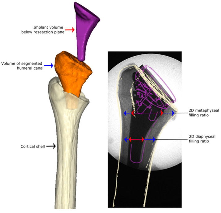
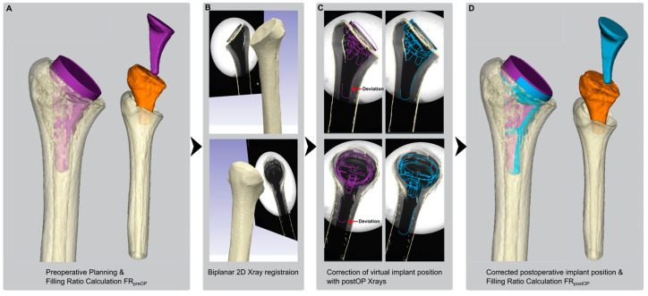
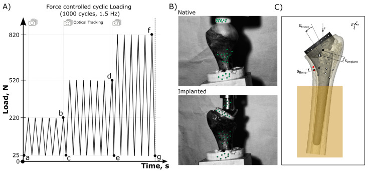
Methods: Forty cadaveric shoulder specimens were analyzed in a clinical computed tomography (CT) scanner allowing for segmentation of the humeral canal to calculate volumetric measures which were verified postoperatively with plain radiographs. Virtual implant positioning allowed for group assignment (VFR < 0.72): Standard stem with low (n = 10) and high (n = 10) filling ratios, a short stem with low (n = 10) and high filling ratios (n = 10). Biomechanical testing included cyclic loading of the native bone and the implanted humeral component. Optical recording allowed for spatial implant tracking and the quantification of cortical bone deformations in the proximal humerus.
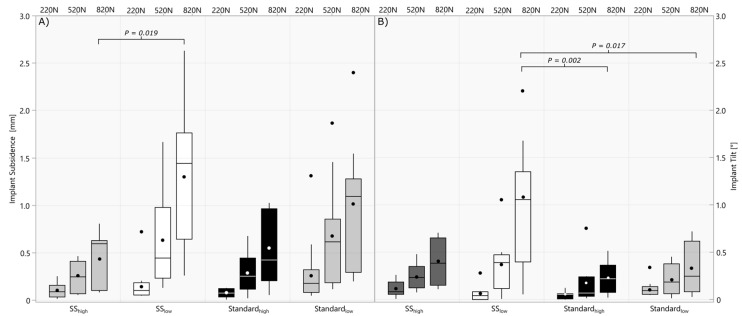
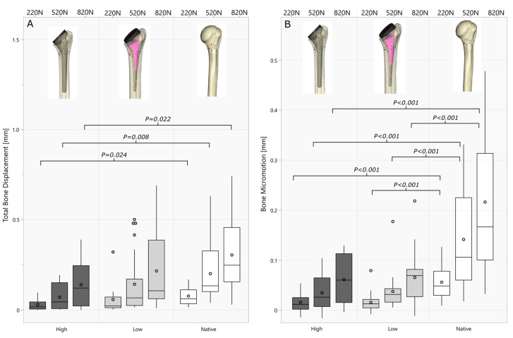
Results: Planned filling ratios based on 3D volumetric measures had a good-to-excellent correlation (ICC = 0.835; p < 0.001) with implanted filling ratios. Lower canal fill ratios resulted in significantly higher variability between short and standard stems regarding implant tilt (820 N: p = 0.030) and subsidence (220 N: p = 0.046, 520 N: p = 0.007 and 820 N: p = 0.005). Higher filling ratios resulted in significantly lower bone deformations in the medial calcar area compared to the native bone, while the bone deformations in lower filling ratios did not differ significantly (p > 0.177).
Conclusions: Lower canal filling ratios maintain dynamic bone loading in the medial calcar of the humerus similar to the native situation in this biomechanical loading setup. Short stems implanted with a low filling ratio have an increased risk for implant tilt and subsidence compared to high filling ratios or standard stems.
Keywords: CT imaging; biomechanics; bone deformation; canal fill; micromotion; reverse shoulder arthroplasty; short stem; standard stem; stress shielding.
Conflict of interest statement
D.R. is an employee of Arthrex. P.R. receives consulting fees and honoraria from Arthrex, receives support for travel to meetings for the study or other purposes from Arthrex. P.D. receives consulting fees and honoraria from Arthrex, receives support for travel to meetings for the study or other purposes from Arthrex, and receives royalties from Arthrex. B.W. receives consulting fees and honoraria from Arthrex, Pacira and Lifenet, receives research support from Arthrex, Zimmer Biomet, Exactech, Pacira and Lifenet and receives support for travel to meetings for the study or other purposes from Arthrex. M.W. declares no conflicts of interests. C.W. is an employee of Arthrex. P.E.M. receives consulting fees and honoraria from BBraun Aesculap and Medacta and the affiliated research institute receives research support from Arthrex. There were no financial payments or other benefits related to the subject of this article. S.B. is an employee of Arthrex.
Figures

References
-
- Esfandiari A., Hamoodi Z., Newton A., Nixon M., Webb M., Kenyon P. The incidence of humeral bone resorption in uncemented reverse shoulder arthroplasty and the impact on functional outcomes. Semin. Arthroplast. JSES. 2022;32:638–643. doi: 10.1053/j.sart.2022.04.009. - DOI
-
- Testa E.J., Albright J.A., Lemme N.J., Molla V., McCrae B., Daniels A.H., Paxton E.S. Increased Risk of Periprosthetic Fractures and Revision Arthroplasty in Patients Undergoing Shoulder Arthroplasty With a History of Prior Fragility Fractures: A Matched Cohort Analysis. J. Am. Acad. Orthop. Surg. 2023;31:e473–e480. doi: 10.5435/JAAOS-D-22-00752. - DOI - PubMed
-
- ASES Complications of RSA Multicenter Research Group. Mahendraraj K.A., Abboud J., Armstrong A., Austin L., Brolin T., Entezari V., Friedman L., Garrigues G.E., Grawe B., et al. Predictors of acromial and scapular stress fracture after reverse shoulder arthroplasty: A study by the ASES Complications of RSA Multicenter Research Group. J. Shoulder Elb. Surg. 2021;30:2296–2305. doi: 10.1016/j.jse.2021.02.008. - DOI - PubMed
LinkOut - more resources
Full Text Sources
