. 2024 Nov 21;14(12):649.
doi: 10.3390/metabo14120649.

Effects of Ammonia Stress on Liver Tissue Structure, Enzyme Activities, and Metabolome of Juvenile Largemouth Bass Micropterus salmoides

Affiliations

Effects of Ammonia Stress on Liver Tissue Structure, Enzyme Activities, and Metabolome of Juvenile Largemouth Bass Micropterus salmoides

Decheng Pu et al. Metabolites. .

Abstract

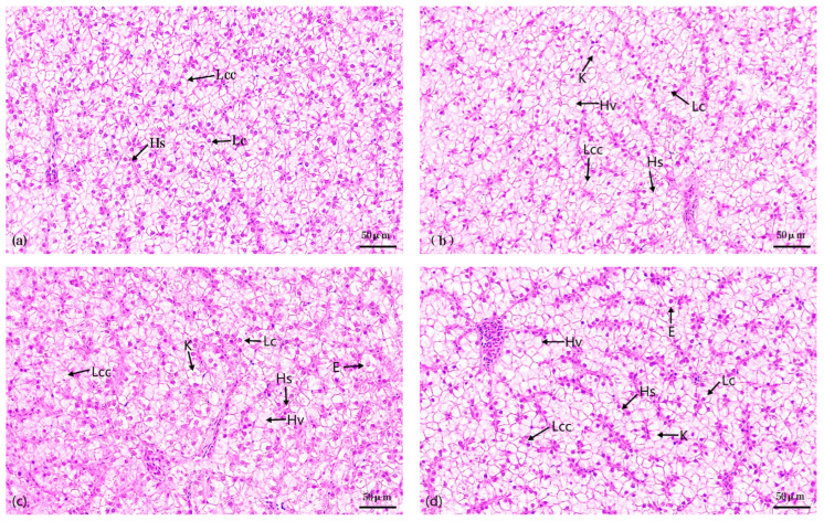
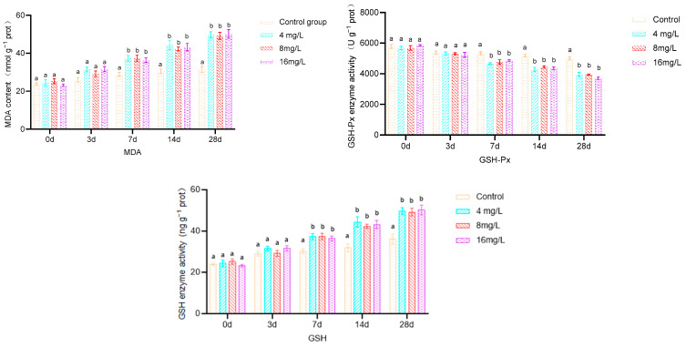
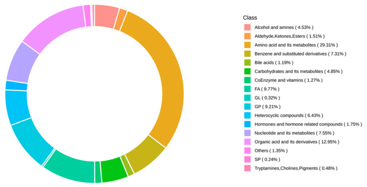
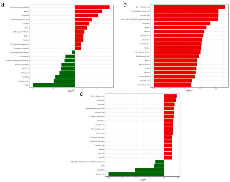
Background: Ammonia, a ubiquitous contaminant in aquatic ecosystems, poses multifaceted threats to fish species at elevated concentrations. Methods: In order to investigate the toxic effects of chronic ammonia stress on the liver of juvenile Micropterus salmoides, the present experiment was conducted to investigate the differences in changes in liver tissue structure, enzyme activities, and metabolomes after 28 days of ammonia exposure (0, 4, 8, and 16 mg/L). Results: The findings revealed that ammonia exposure induced significant oxidative stress in the liver, manifesting in decreased activities of antioxidant enzymes SOD and GSH-Px, elevated levels of GSH, GST, and MDA, and heightened activities of immune enzymes LZM, ALP, and ACP. An increase in ammonia concentration exacerbated liver tissue damage. Metabolome analysis further unveiled perturbations in liver metabolites of Micropterus salmoides exposed to ammonia, with Ala-His emerging as a potentially pivotal functional substance under chronic stress. Specifically, the 4 mg/L group responded to ammonia toxicity by augmenting GSH and L-Carnosine levels, the 8 mg/L group detoxified via upregulation of L-Glutamine, and the 16 mg/L group mitigated toxicity through the urea synthesis pathway. Conclusions: This research offers preliminary insights into the toxicological responses of Micropterus salmoides under chronic ammonia stress. It is suggested that the duration of ammonia concentration exceeding 4 mg/L in high-density aquaculture should not exceed 7 days.

Keywords: ammonia toxicity; immune enzymes; metabolites; oxidative stress; tissue damage.


Conflict of interest statement

The authors declare no conflicts of interest.

Figures

Figure 1
Figure 1
Effects of 28 d exposure to different ammonia concentrations on the liver tissue structure of Micropterus salmoides. Note: (a). control group; (b). 4 mg/L treatment group; (c). 8 mg/L treatment group; (d). 16 mg/L treatment group; Lc. hepatocytes; Hs. hepatic sinusoids; Lcc. hepatocyte cords; K. hepatocyte nucleolysis; Hv. hepatocyte vacuolation; E. hepatocyte enlargement.
Figure 2
Figure 2
Effect of 28 d exposure to different ammonia concentrations on liver antioxidant enzyme activities of Micropterus salmoides. Note: Different letters indicate significant differences between groups with different ammonia concentrations at the same time (p < 0.05); same below.
Figure 2
Figure 2
Effect of 28 d exposure to different ammonia concentrations on liver antioxidant enzyme activities of Micropterus salmoides. Note: Different letters indicate significant differences between groups with different ammonia concentrations at the same time (p < 0.05); same below.
Figure 3
Figure 3
Effect of 28 d of exposure to different ammonia concentrations on liver immunoenzyme activities of Micropterus salmoides.
Figure 4
Figure 4
Ring diagram of metabolite class composition. Note: Each color represents a metabolite class, and the area of the color block indicates the percentage of that class that is represented.
Figure 5
Figure 5
Map of OPLS-DA scores. (a), LIV−A1_vs._LIV−C; (b), LIV−A2_vs._LIV−C; (c), LIV−A3_vs._LIV−C. Note: the horizontal coordinate indicates the predicted principal component and the horizontal direction shows the gap between groups; the vertical coordinate indicates the orthogonal principal component and the vertical direction shows the gap within groups; and the percentage indicates the explanation rate of this component to the dataset. Each point in the graph represents a sample, samples in the same group are represented using the same color, and the Group is the subgroup.
Figure 6
Figure 6
Volcano map of differential metabolites. (a), LIV−A1_vs._LIV−C; (b), LIV−A2_vs._LIV−C; (c), LIV−A3_vs._LIV−C. Note: each dot in the volcano diagram represents a metabolite, where green dots represent downregulated differential metabolites, red dots represent upregulated differential metabolites, and grey dots represent metabolites detected but with insignificant differences. The horizontal coordinate represents the logarithm of the multiplicity of the differences in the relative content of a metabolite between the two groups of samples (log2FC), and the greater the absolute value of the horizontal coordinate, the greater the difference of relative content of the substance between the two groups of samples. VIP + FC + p-value screening condition: the vertical coordinate indicates the significance level of difference (−log10p-value), and the size of the dot represents the VIP value.
Figure 7
Figure 7
Variance multiplier bar graph. (a), LIV−A1_vs._LIV−C; (b), LIV−A2_vs._LIV−C; (c), LIV−A3_vs._LIV−C. Note: the horizontal coordinate is the log2FC of the differential metabolite, i.e., the value of the multiplicity of the difference of the differential metabolite taken logarithmically with 2 as the base, and the vertical coordinate is the differential metabolite. Red color represents upregulation of metabolite content and green color represents downregulation of metabolite content.
Figure 8
Figure 8
Venn diagram of differential metabolites in different comparison groups. Note: each circle in the figure represents a comparison group, the number of overlapping circles represents the number of differential metabolites common to the comparison groups, and the number with no overlap represents the number of differential metabolites specific to the comparison groups.
Figure 9
Figure 9
KEGG pathway of differential metabolites in the liver of Micropterus salmoides enriched in the top 20. (a), LIV−A1_vs._LIV−C; (b), LIV−A2_vs._LIV−C; (c), LIV−A3_vs._LIV−C. Note: the horizontal coordinate indicates the Rich Factor corresponding to each pathway. The vertical coordinate is the pathway name (sorted by p-value), and the color of the dots reflects the size of the p-value; the redder the more significant the enrichment. The size of the dots represents the number of different metabolites enriched.
Figure 10
Figure 10
Heatmap of differential metabolite clustering of the KEGG pathway. (a), LIV−A1_vs._LIV−C; (b), LIV−A2_vs._LIV−C; (c), LIV−A3_vs._LIV−C. Note: the horizontal coordinate is the sample name. The vertical coordinate is the differential metabolite, different colors are the colors filled with different values obtained after the standardized treatment of different relative contents (red represents high content, green represents low content). The comment bar above the heat map corresponds to the grouping of the samples (Group). The dendrogram on the left side of the heat map represents the results of the hierarchical clustering of the differential metabolites. The comment bar on the right side of the clustering tree corresponds to the substance class (Class), and different colors represent different substance classes. The annotation bars on the right side of the clustering tree correspond to the first class of substances, and different colors represent different substance classes.

Similar articles

Cited by

References

    1. Liang Z., Li R., Zhao D., Wang L., Sun M. Ammonia exposure induces oxidative stress, endoplasmic reticulum stress and apoptosis in hepatopancreas of pacific white shrimp (Litopenaeus vannamei) Fish Shellfish Immunol. 2016;54:523–528. doi: 10.1016/j.fsi.2016.05.009. - DOI - PubMed
    1. Zhang T., Zhou Y., Wen H., Ma X., Xu D. Integrated analysis of physiological, transcriptome, and metabolome analyses of the gills in Solenaia oleivora under ammonia exposure. Ecotoxicol. Environ. Saf. 2024;271:115949. doi: 10.1016/j.ecoenv.2024.115949. - DOI - PubMed
    1. Wang F., Huang L., Liao M., Dong W., Liu C., Zhuang X., Liu Y., Yin X., Liang Q., Wang W. Pva-miR-252 participates in ammonia nitrogen-induced oxidative stress by modulating autophagy in Penaeus vannamei. Ecotoxicol. Environ. Saf. 2021;225:112774. doi: 10.1016/j.ecoenv.2021.112774. - DOI - PubMed
    1. Zhang W., Xia S., Zhu J., Miao L., Ren M., Lin Y., Ge X., Sun S. Growth performance, physiological response and histology changes of juvenile blunt snout bream, Megalobrama amblycephala exposed to chronic ammonia. Aquaculture. 2019;506:424–436. doi: 10.1016/j.aquaculture.2019.03.072. - DOI
    1. Sun Z., Wang S., Zhang M., Jiang H., Li M. Chronic toxicity study of ammonia exposure in juvenile yellow catfish Pelteobagrus fulvidraco. Aquaculture. 2023;567:739266. doi: 10.1016/j.aquaculture.2023.739266. - DOI

LinkOut - more resources