Review
. 2024 Nov 25;14(12):659.
doi: 10.3390/metabo14120659.

Structural and Functional Integration of Tissue-Nonspecific Alkaline Phosphatase Within the Alkaline Phosphatase Superfamily: Evolutionary Insights and Functional Implications

Affiliations
Review

Structural and Functional Integration of Tissue-Nonspecific Alkaline Phosphatase Within the Alkaline Phosphatase Superfamily: Evolutionary Insights and Functional Implications

Iliass Imam et al. Metabolites. .

Abstract

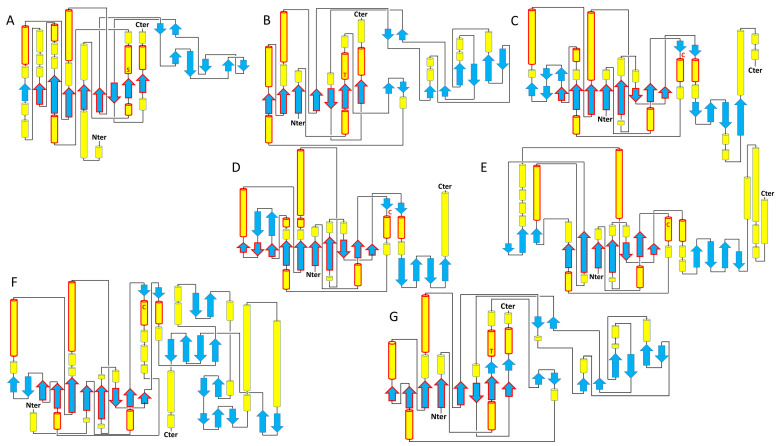
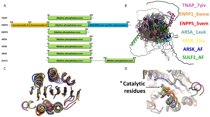
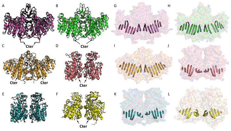
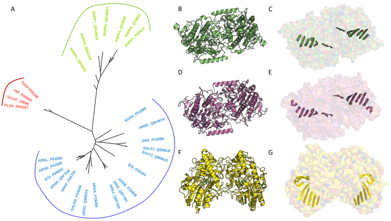
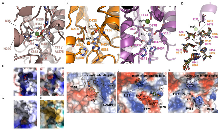
Phosphatases are enzymes that catalyze the hydrolysis of phosphate esters. They play critical roles in diverse biological processes such as extracellular nucleotide homeostasis, transport of molecules across membranes, intracellular signaling pathways, or vertebrate mineralization. Among them, tissue-nonspecific alkaline phosphatase (TNAP) is today increasingly studied, due to its ubiquitous expression and its ability to dephosphorylate a very broad range of substrates and participate in several different biological functions. For instance, TNAP hydrolyzes inorganic pyrophosphate (PPi) to allow skeletal and dental mineralization. Additionally, TNAP hydrolyzes pyridoxal phosphate to allow cellular pyridoxal uptake, and stimulate vitamin B6-dependent reactions. Furthermore, TNAP has been identified as a key enzyme in non-shivering adaptive thermogenesis, by dephosphorylating phosphocreatine in the mitochondrial creatine futile cycle. This latter recent discovery and others suggest that the list of substrates and functions of TNAP may be much longer than previously thought. In the present review, we sought to examine TNAP within the alkaline phosphatase (AP) superfamily, comparing its sequence, structure, and evolutionary trajectory. The AP superfamily, characterized by a conserved central folding motif of a mixed beta-sheet flanked by alpha-helices, includes six subfamilies: AP, arylsulfatases (ARS), ectonucleotide pyrophosphatases/phosphodiesterases (ENPP), phosphoglycerate mutases (PGM), phosphonoacetate hydrolases, and phosphopentomutases. Interestingly, TNAP and several ENPP family members appear to participate in the same metabolic pathways and functions. For instance, extra-skeletal mineralization in vertebrates is inhibited by ENPP1-mediated ATP hydrolysis into the mineralization inhibitor PPi, which is hydrolyzed by TNAP expressed in the skeleton. Better understanding how TNAP and other AP family members differ structurally will be very useful to clarify their complementary functions. Structurally, TNAP shares the conserved catalytic core with other AP superfamily members but has unique features affecting substrate specificity and activity. The review also aims to highlight the importance of oligomerization in enzyme stability and function, and the role of conserved metal ion coordination, particularly magnesium, in APs. By exploring the structural and functional diversity within the AP superfamily, and discussing to which extent its members exert redundant, complementary, or specific functions, this review illuminates the evolutionary pressures shaping these enzymes and their broad physiological roles, offering insights into TNAP's multifunctionality and its implications for health and disease.

Keywords: alkaline phosphatase (AP) superfamily; arylsulfatases (ARS); ectonucleotide pyrophosphatases/phosphodiesterases (ENPP); tissue-nonspecific alkaline phosphatase (TNAP).


Conflict of interest statement

The authors declare no conflicts of interest.

Figures

Figure 1
Figure 1
Topology of representative members of the AP superfamily. (A) Human TNAP, (B) human ENPP5, (C) human ARSA, (D) human ARSB, (E) human ARSK, (F) human SULF1, (G) human ENPP1. The central core is conserved in members of the AP, ENPP, and ARS families, with a minimal conservation of 7 to 8 beta strands and 6 to 7 alpha helices highlighted in solid red lines. The additional domains of ENPP1, somatomedin at the N-terminus (1–192), and non-specific DNA/RNA endonuclease at the C-terminus (637–925) are not shown for clarity. The secondary structures, alpha helices, and beta strands are shown in yellow and blue, respectively. The catalytic residues are indicated by the letters S (serine), T (threonine), and C (cysteine).
Figure 2
Figure 2
Conservation of the core in the AP superfamily. (A) Examples of domain organization in various members of the superfamily. (B) Superposition of 7 representative members of the superfamily, including AP, ENPP, and ARS. The members are TNAP (ALPL) in pink (PDB code: 7YIV), ENPP1 in orange (PDB code: 6WEW), ENPP5 in red (PDB code: 5VEM), ARSA in light blue (PDB code: 1AUK), ARSB in yellow (PDB code: 1FSU), ARSK in dark blue (AlphaFold model), and SULF1 in light green (AlphaFold model). The superposition focuses solely on the central core, highlighting the extreme organizational variability and the lack of overall folding conservation outside the catalytic core. (C) Extraction of the minimal secondary structures from the previous superposition that constitutes the central core. This reveals the high conservation of the central core’s folding, with its 7 to 8 beta strands and 6 to 7 alpha helices. The star marks the position of the nucleophilic catalytic residue: serine for TNAP, threonine for ENPP1 and ENPP5, modified cysteine for ARSA (formylglycine) and ARSB (sulfate ester), and cysteine for the AlphaFold models of ARSK and SULF1. (D) Conserved positioning of the catalytic residue at the end of the helix.
Figure 3
Figure 3
Comparison of dimeric organization in AP and ENPP. (AF) Examples of characteristic dimers of AP and ENPP. (GL) Organization of the central β-sheet characteristic of the AP family. The Van der Waals surface of the dimer is shown in transparency. The dimeric organization is consistent across the AP family, including human enzymes like TNAP (PDB code: 7YIV) (panels A,G) and PLAP (PDB code: 1ZED) (panels B,H), as well as the E. coli AP enzyme (panels C,E). This organization features conserved central β-sheets, typical of the AP fold, facing each other without complementary interactions (panels G,H,E). A similar AP-like interface is observed in some ENPP family members, particularly in murine ENPP6 (panel D) (PDB code: 5EGH). An AlphaFold model of the human ENPP6 dimer suggests a similar organization (panel F) (PDB code: 5VEN). The same AP-like interface is observed in murine ENPP5, with the conserved central β-sheets facing each other with a slight offset and no complementary interactions (panels J,K,L).
Figure 4
Figure 4
Evolution of members of the AP superfamily: (A) Phylogenetic tree of the AP superfamily members. Multiple sequence alignment of all human members of the AP superfamily was performed with the MAFFT program version 7 [54]. The sequences used were retrieved from the UniProtKB database, with the corresponding UniProt codes listed next to the enzyme names. Conserved blocks were selected by using BMGE1.12 [55] and the BLOSUM30 [56] matrix. Two hundred sites were kept for further analysis after character trimming was performed by BMGE [55]. Phylogenetic analyses were performed, with the LG model and a gamma correction, using a bootstrapped maximum-likelihood approach with PhyML 3.0 [57]. The phylogenetic tree was generated and visualized with iTOL software Version 6.9.1 [58]. (B,D,F) Examples of characteristic dimers observed in ARS. (C,E,G) Central β-sheet illustrating the diversity of organization in ARS. The Van der Waals surface of the dimer is shown in transparency. Two types of organization are presented here: one corresponding to GALNS (PDB code: 4FDJ) and ARSA (PDB code: 1AUK) (BE) and the other to N-sulphoglucosamine sulphohydrolase (SGSH; PDB code: 4MHX) (F,G).
Figure 5
Figure 5
Comparison of the catalytic centers of AP, ENPP, and ARS, representative of each family. (A) Catalytic center of ARSC (PDB code: 8EG3). Conserved residues of the family are indicated. Calcium is shown as a green sphere. (B) Catalytic center of ENPP1 (PDB code: 6WEU). Conserved residues of the family are indicated. Zinc 1 and 2 are shown as orange spheres. (C) Catalytic center of TNAP (PDB code: 7YIV). Conserved residues of the family are indicated. Zinc 1 and 2 are shown as pink spheres, and magnesium is shown as a green sphere. (D) Superposition of representative members from the AP and ENPP families. Conserved residues between the two families are shown, indicating that the catalytic residue and the residues interacting with zinc are almost perfectly superimposable. The residues of TNAP and ENPP1 are numbered. The structures used in the comparison are TNAP in purple (PDB code: 7YIV), PLAP in light green (PDB code: 1ZED), ENPP1 in orange (PDB code: 6WEU), ENPP2 in yellow (PDB code: 5M7M), ENPP3 in light blue (PDB code: 6C02), ENPP4 in cyan (PDB code: 4LQY), ENPP5 in red (PDB code: 5VEM), and ENPP7 in dark green (PDB code: 5UDY). (EH) Characteristic crevice of ARS with the catalytic pocket at its bottom. (E,F) Electrostatic surfaces of GALNS (PDB code: 4FDJ) and IDS (PDB code: 5FQL) reveal a highly positively charged crevice. A citrate molecule is visible at the bottom of the GALNS structure. (G,H) Electrostatic and hydrophobic surfaces of the binding site of ARSC (PDB code: 8EG3), showing one side that is positively charged (blue) (G) and the other side that is hydrophobic (yellow) (H). (IK) Electrostatic surfaces of ENPP1, PLAP, and TNAP, with all three enzymes similarly oriented. Zn2+ ions are indicated by red and gray spheres. In ENPP1, adenosine-5′-thio-monophosphate is visible in the active site, with the base positioned in the nucleotide-binding slot stabilized by residues Y340 and F257 (I). In PLAP and TNAP, the magnesium-binding helix occupies the nucleotide-binding slot present in ENPP1, necessitating a different substrate accommodation. This is observed in PLAP (J) with the presence of PNP. The gatekeeper residues, D428 and E429 for PLAP and R450 and H451 for TNAP, are shown as sticks (J,K).

Similar articles

Cited by

References

    1. Murshed M., Harmey D., Millán J.L., McKee M.D., Karsenty G. Unique coexpression in osteoblasts of broadly expressed genes accounts for the spatial restriction of ECM mineralization to bone. Genes Dev. 2005;19:1093–1104. doi: 10.1101/gad.1276205. - DOI - PMC - PubMed
    1. Whyte M.P., Fujita K.P., Moseley S., Thompson D.D., McAlister W.H. Validation of a Novel Scoring System for Changes in Skeletal Manifestations of Hypophosphatasia in Newborns, Infants, and Children: The Radiographic Global Impression of Change Scale. J. Bone Miner. Res. 2018;33:868–874. doi: 10.1002/jbmr.3377. - DOI - PubMed
    1. Fedde K.N., Blair L., Silverstein J., Coburn S.P., Ryan L.M., Weinstein R.S., Waymire K., Narisawa S., Millán J.L., Macgregor G.R., et al. Alkaline phosphatase knock-out mice recapitulate the metabolic and skeletal defects of infantile hypophosphatasia. J. Bone Miner. Res. 1999;14:2015–2026. doi: 10.1359/jbmr.1999.14.12.2015. - DOI - PMC - PubMed
    1. Schippers M., Post E., Eichhorn I., Langeland J., Beljaars L., Malo M.S., Hodin R.A., Millán J.L., Popov Y., Schuppan D., et al. Phosphate Groups in the Lipid A Moiety Determine the Effects of LPS on Hepatic Stellate Cells: A Role for LPS-Dephosphorylating Activity in Liver Fibrosis. Cells. 2020;9:2708. doi: 10.3390/cells9122708. - DOI - PMC - PubMed
    1. Bessueille L., Briolay A., Como J., Mebarek S., Mansouri C., Gleizes M., El Jamal A., Buchet R., Dumontet C., Matera E., et al. Tissue-nonspecific alkaline phosphatase is an anti-inflammatory nucleotidase. Bone. 2020;133:115262. doi: 10.1016/j.bone.2020.115262. - DOI - PMC - PubMed

LinkOut - more resources