Review
. 2024 Dec 22;14(12):723.
doi: 10.3390/metabo14120723.

Circadian Influences on Brain Lipid Metabolism and Neurodegenerative Diseases

Affiliations
Review

Circadian Influences on Brain Lipid Metabolism and Neurodegenerative Diseases

Yusuf Hussain et al. Metabolites. .

Abstract

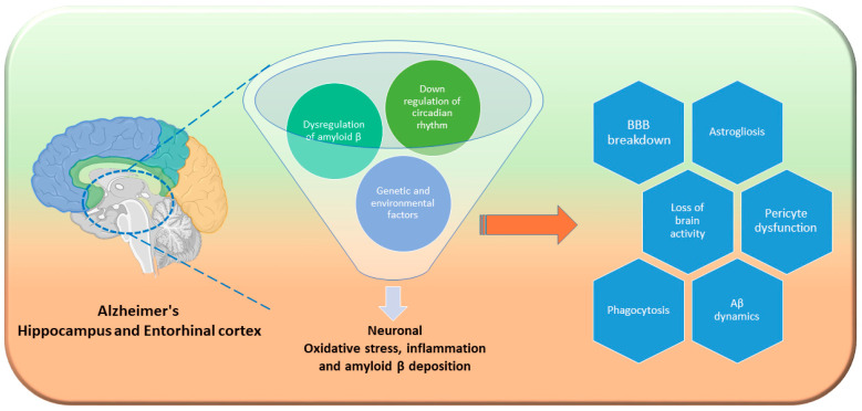
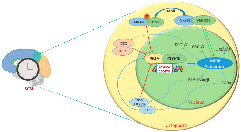
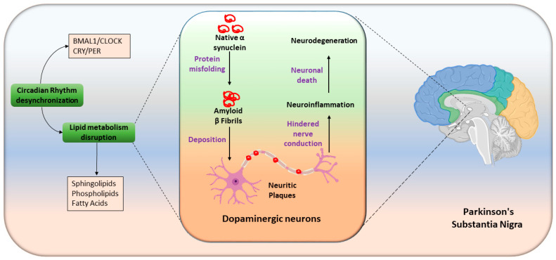
Circadian rhythms are intrinsic, 24 h cycles that regulate key physiological, mental, and behavioral processes, including sleep-wake cycles, hormone secretion, and metabolism. These rhythms are controlled by the brain's suprachiasmatic nucleus, which synchronizes with environmental signals, such as light and temperature, and consequently maintains alignment with the day-night cycle. Molecular feedback loops, driven by core circadian "clock genes", such as Clock, Bmal1, Per, and Cry, are essential for rhythmic gene expression; disruptions in these feedback loops are associated with various health issues. Dysregulated lipid metabolism in the brain has been implicated in the pathogenesis of neurological disorders by contributing to oxidative stress, neuroinflammation, and synaptic dysfunction, as observed in conditions such as Alzheimer's and Parkinson's diseases. Disruptions in circadian gene expression have been shown to perturb lipid regulatory mechanisms in the brain, thereby triggering neuroinflammatory responses and oxidative damage. This review synthesizes current insights into the interconnections between circadian rhythms and lipid metabolism, with a focus on their roles in neurological health and disease. It further examines how the desynchronization of circadian genes affects lipid metabolism and explores the potential mechanisms through which disrupted circadian signaling might contribute to the pathophysiology of neurodegenerative disorders.

Keywords: Alzheimer’s disease; brain function; cholesterol; circadian rhythm; fatty acid; lipid metabolism.


Conflict of interest statement

The authors declare no conflicts of interest.

Figures

Figure 4
Figure 4
Hippocampal and entorhinal cortex insult in Alzheimer’s disease neurodegeneration. Primarily, the hippocampus and entorhinal cortex two are key regions for learning, memory, and spatial orientation. Genetic and environmental factors compromise circadian rhythm, followed by lipid metabolism. Loss of central circadian rhythms disrupts amyloid β fluid oscillations, speeding up amyloid plaque buildup. For example, downregulation of Bmal1 in the brain parenchyma elevates ApoE expression and encourages fibrillar plaque buildup [55]. Combined with Aβ plaque buildup and tau tangles, circadian disruption worsens neuroinflammation and impairs synaptic plasticity [212]. Subsequently, abnormal autophagy leads to BBB breakdown, and a loss of pericyte functioning aggravates the condition [215]. As circadian rhythm disturbances compound this molecular damage, neurodegeneration accelerates and drives AD progression.
Figure 1
Figure 1
Circadian clock gene regulation in brain. The central circadian clock in the SCN regulates body rhythms and sends signals to peripheral clocks. The CLOCK/BMAL1 complex binds E-box elements on target genes (CRY1/2, PER1/2/3, REV-ERBα/β, and RORα). PER and CRY proteins form cytoplasmic heterodimers that shuttle to the nucleus. After phosphorylation by CK1δ and CK1ε, PER/CRY suppress E-box gene transcription via CLOCK/BMAL1.
Figure 2
Figure 2
Lipid trafficking in the brain. Astrocytes synthesize cholesterol via HMG-CoA reductase (HMG-CoAR), package it into ApoE lipoproteins, and export it via ABCA1. Neurons receive APOE/C1 lipoprotein for neurite growth, synaptogenesis, or conversion to 24-OHC by cholesterol 24-hydroxylase (CYP46). Stimulated or stressed neurons release FAs in APOE particle lipoprotein to astrocytes for degradation or storage in lipid droplets. Lipids for oligodendrocyte myelination/remyelination are synthesized by both astrocytes and oligodendrocytes, and ApoE aids in astrocyte-to-oligodendrocyte transport. Also, neurons can transfer cholesterol-rich lipoproteins to oligodendrocytes through low-density lipoprotein receptor (LDLr) on oligodendrocytes. Excess saturated FAs from astrocytes can cause oligodendrocyte death via lipoapoptosis. Lipids from myelin debris activate triggering receptor expressed on myeloid cells 2 (also called TREM2) signaling. TREM2 expression triggers spleen tyrosine kinase (Syk) activation, triggering lipid metabolism genes that aid in lipid droplet breakdown and lipid efflux, and is also known to participate in neurological disorders [97,98,99,100,101].
Figure 3
Figure 3
Dysregulated circadian rhythm and lipid metabolism drive neuronal insult in Huntington’s disease. Environmental factors downregulate circadian genes, thereby disrupting neuronal lipid homeostasis. This disturbance promotes increased lipid uptake by neurons, and results in lipid accumulation and subsequent downregulation of ABC transporters, including ABCA1, ABCA4, and ABCG5, which are critical for phospholipid and cholesterol transport. Diminished ABC transporter activity exacerbates lipid aggregation, impairs lipophagy, and hinders the clearance of excess lipids. This lipid overload contributes to septin dysregulation, which in turn impairs nerve conduction, and ultimately triggers neuroinflammation and neuronal degeneration associated with HD.
Figure 5
Figure 5
Pathophysiology of Parkinson’s disease: Under physiological conditions, α-synuclein exists as a soluble random coil. Pathological conditions are caused by abnormal lipid metabolism and desynchronized circadian rhythm. α-synuclein is misfolded, thus forming toxic dimers, trimers, and oligomers. These misfolded forms aggregate into protofibrils, intermediates, and amyloid fibrils. Aggregates form Lewy bodies or Lewy neurites and deposited over dopaminergic neurons and lead to neuroinflammation, neuronal death, and subsequently degradation of the substabtia nigra.

Similar articles

References

    1. Bass J. Interorgan rhythmicity as a feature of healthful metabolism. Cell Metab. 2024;36:655–669. doi: 10.1016/j.cmet.2024.01.009. - DOI - PMC - PubMed
    1. Allada R., Bass J. Circadian Mechanisms in Medicine. N. Engl. J. Med. 2021;384:550–561. doi: 10.1056/NEJMra1802337. - DOI - PMC - PubMed
    1. Walker W.H., 2nd, Walton J.C., DeVries A.C., Nelson R.J. Circadian rhythm disruption and mental health. Transl. Psychiatry. 2020;10:28. doi: 10.1038/s41398-020-0694-0. - DOI - PMC - PubMed
    1. Boivin D.B., Boudreau P., Kosmadopoulos A. Disturbance of the Circadian System in Shift Work and Its Health Impact. J. Biol. Rhythms. 2022;37:3–28. doi: 10.1177/07487304211064218. - DOI - PMC - PubMed
    1. Fagiani F., Di Marino D., Romagnoli A., Travelli C., Voltan D., Di Cesare Mannelli L., Racchi M., Govoni S., Lanni C. Molecular regulations of circadian rhythm and implications for physiology and diseases. Signal Transduct. Target. Ther. 2022;7:41. doi: 10.1038/s41392-022-00899-y. - DOI - PMC - PubMed

LinkOut - more resources