

Placental Macrovascular Pattern from Pregnancies with Maternal Hypertensive and Fetal Growth Capacity Complications
- PMID: 39728685
- PMCID: PMC11679333
- DOI: 10.3390/pathophysiology31040050
Placental Macrovascular Pattern from Pregnancies with Maternal Hypertensive and Fetal Growth Capacity Complications
Abstract
Histomorphometric measurements of the wall thickness and internal diameter of the macrovessels of the chorionic villi of placentas from pregnancies complicated by preeclampsia or fetal growth restriction in comparison with normotensive pregnancy.
Methods: The research included placentas from singleton pregnancies complicated by preeclampsia and/or fetal growth restriction, women delivered in medical institutions in Karaganda city (Kazakhstan). Placentas were divided into three groups: PE (n = 59), isolated FGR (n = 24), and PE with FGR (n = 41). The control group consisted of normotensive pregnancies, compared by gestation period. Placental examination and selection of placental tissue fragments were carried out in accordance with the consensus recommendations of the Amsterdam Placental Workshop Group. The sections were stained with hematoxylin and eosin and Masson trichrome. Morphometric measurements were performed using ImageJ software version 1.52p.
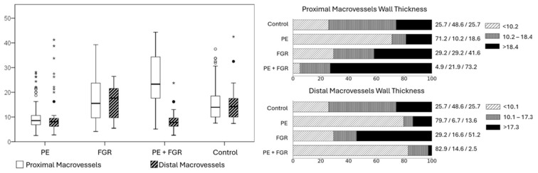
Results: Our data showed that, in the PE group, there was a significant decrease in the wall thickness of the proximal and distal vessels with an increase in internal diameter compared with the control group (p < 0.01). In the PE + FGR group, there was a thickening of the wall of the proximal part of the vessels with a decrease in their lumen and a decrease in the wall thickness of the vessels with an increase in the lumen in the distal part compared with the control group (p < 0.01).
Conclusions: Two histopatterns of placental macrovessels in preeclampsia were revealed: the histophenotype of diffuse (proximal and distal) ectatic macroangiopathy with a thin vascular wall with a decrease in the thickness of the muscle layer and the histophenotype of proximal fibromuscular sclerosis with vascular obliteration/spasm and distal ectatic macroangiopathy. We believe that significant structural differences in vascular remodeling may reflect the different temporal and spatial nature of the pathological factor. Future research is needed to investigate the associations between histopatterns of placental vascular remodeling in preeclampsia and long-term perinatal/maternal outcomes.
Keywords: chorionic villi vessels; fetal growth restriction; morphometric; placenta; preeclampsia.
Conflict of interest statement
The authors declare no conflicts of interest.
Figures
 ) minor emissions, defined as values in the range from 1.5 to 3 interquartile ranges (IQR); stars (
 )—extreme emissions, defined as values greater than 3 interquartile ranges (IQR).); Assessment of placental macrovascular wall thickness in the preeclampsia and fetal growth restriction groups compared with the control group.
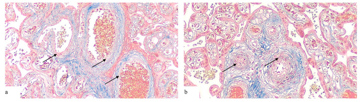
References
-
- Brain K.L., Allison B.J., Niu Y., Cross C.M., Itani N., Kane A.D., Herrera E.A., Skeffington K.L., Botting K.J., Giussani D.A. Intervention against hypertension in the next generation programmed by developmental hypoxia. PLoS Biol. 2019;17:e2006552. doi: 10.1371/journal.pbio.2006552. - DOI - PMC - PubMed
-
- Chen X., Qi L., Fan X., Tao H., Zhang M., Gao Q., Liu Y., Xu T., Zhang P., Su H., et al. Prenatal hypoxia affected endothelium-dependent vasodilation in mesenteric arteries of aged offspring via increased oxidative stress. Hypertens. Res. 2019;42:863–875. doi: 10.1038/s41440-018-0181-7. - DOI - PubMed
-
- Hula N., Spaans F., Vu J., Quon A., Kirschenman R., Cooke C.M., Phillips T.J., Case C.P., Davidge S.T. Placental treatment improves cardiac tolerance to ischemia/reperfusion insult in adult male and female offspring exposed to prenatal hypoxia. Pharmacol. Res. 2021;165:105461. doi: 10.1016/j.phrs.2021.105461. - DOI - PubMed
LinkOut - more resources
Full Text Sources
Miscellaneous