. 2024 Dec 2;65(14):41.
doi: 10.1167/iovs.65.14.41.

Functional Roles of Pigment Epithelium-Derived Factor in Retinal Degenerative and Vascular Disorders: A Scoping Review

Affiliations

Functional Roles of Pigment Epithelium-Derived Factor in Retinal Degenerative and Vascular Disorders: A Scoping Review

Thomas Stax Jakobsen et al. Invest Ophthalmol Vis Sci. .

Abstract

Purpose: This review explores the role of pigment epithelium-derived factor (PEDF) in retinal degenerative and vascular disorders and assesses its potential both as an adjunct to established vascular endothelial growth factor inhibiting treatments for retinal vascular diseases and as a neuroprotective therapeutic agent.

Methods: A comprehensive literature review was conducted, focusing on the neuroprotective and anti-angiogenic properties of PEDF. The review evaluated its effects on retinal health, its dysregulation in ocular disorders, and its therapeutic application in preclinical models. Advances in drug delivery, including gene therapy, were also examined.

Results: PEDF, initially identified for promoting neuronal differentiation, is also a potent endogenous angiogenesis inhibitor. Strong anti-angiogenic and neuroprotective effects are observed in preclinical studies. It has pro-apoptotic and antiproliferative effects on endothelial cells thereby reducing neovascularization. Although promising, clinical development is limited with only a single conducted phase I clinical trial for macular neovascularization. Development of PEDF-derived peptides enhances potency and specificity, and emerging gene therapy approaches offer sustained PEDF expression for long-term treatment. However, questions regarding dosage, durability, and efficacy remain, particularly in large animal models.

Conclusions: PEDF shows significant therapeutic potential in preclinical models of retinal degeneration and vascular disorders. Despite inconclusive evidence on PEDF downregulation as a primary disease driver, many studies highlight its therapeutic benefits and favorable safety profile. Advances in gene therapy could enable long-acting PEDF-based treatments, but further research is needed to optimize dosage and durability, potentially leading to clinical trials and expanding treatment options for retinal disorders.


Conflict of interest statement

Disclosure: T.S. Jakobsen, None; R.L. Adsersen, None; A.L. Askou, None; T.J. Corydon, None

Figures

Figure 1.
Figure 1.
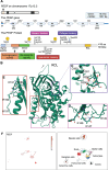
Schematic diagram of the genomic localization and structure of PEDF, the PEDF polypeptide, and PEDF expression in scRNA-seq data from human retina. (A) PEDF is encoded by the PEDF gene located on the forward strand of chromosome 17p13.3 (ID ENST00000254722.9). Exons 1 to 8 are denoted by blue boxes. UTRs (exon 1, and part of exon 2 and 8) are represented by dark blue boxes. The size (in bp) of each element (drawn to scale unless otherwise indicated by backslashes) is indicated by numbers. Numbers in parenthesis indicate translated bases of exons 2 and 8. The size of the mRNA without UTRs is 1257 bp. Introns are indicated by lines. The PEDF protein can be phosphorylated on serine 24, 114, and 227 (indicated by yellow circles), and glycosylated on asparagine 285 (sugar molecule indicated by blue squares and green circles). Two peptide regions contain specific activities: a 34 aa sequence (Asp44-Asn77) responsible for the antiangiogenic activities, and a 44 sequence (Val78-Thr121) responsible for the neurotrophic activities. The RCL as well as the three regions (marked with magenta, red, and purple squares) important for binding of heparin, hyaluronan, or collagen are indicated. (B) Ribbon diagram of human PEDF based on the 2.85 A crystal structure of PEDF (PDB DOI:https://doi.org/10.2210/pdb1IMV/pdb). The folded protein conformation is globular with the RCL (orientation indicated by an arrow) being exposed. It contains 3 β-sheets and 10 α-helices, and an asymmetrical charge distribution, resulting in binding to different components of the ECM. (C) Enlargement of the region containing the acidic amino acids, aspartic acid 256, aspartic acid 258, and aspartic acid 300 (D256, D258, and D300) involved in the collagen binding. The region is rotated approximately 180 degrees compared to B. Isolysine 301 (I301) is shown for orientation purposes. (D) Magnification of the heparin-binding region showing the involved basic amino acids, lysine 146, lysine 147, and arginine 149 (K146, K147, and R149). (E) Expansion of the region holding the basic amino acids, lysine 189, lysine 191, arginine 194, and lysine 197 (K189, K191, R194, and K197) involved in binding to hyaluronan. (F) Normalized PEDF expression in human retinal scRNA-seq data from human retinal tissue (left) with annotated subclusters (right). Expression is evident in a range of cell types particularly prominent in photoreceptor subclusters. (Fig. 1A was created using Biorender.com. Figs. 1B to 1E were prepared by using the 3D structure viewer provided by P.D.B. Fig. 1F processed scRNA-seq data from Lukowski et al. was obtained through the Human Cell Atlas Data Portal [WongAdultRetina]. Analyses were performed using Automated Single Cell Analysis Platform VI. The data were normalized using Seurat and dimensional reduction performed with UMAP. Clusters of cells were identified using Seurat and manually annotated using cell-type marker genes from Lukowski et al.204) PEDF, pigment epithelium-derived factor; aa, amino acids; RCL, reactive center loop; bp, base pairs; kb, kilobases; kDa, kilodaltons; UTR, untranslated region; ECM, extracellular matrix; E, exon.
Figure 2.
Figure 2.
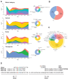
Graphical summaries of PEDF in vivo studies. (A) Trends and distribution of used retinal model categories, delivery vehicles, delivery route, and therapeutic (top to bottom). The color is defined on the corresponding pie chart. For the trend plots the number of studies were binned into 3-year intervals. Model categories include diabetes mellitus, induced barrier disruption, ischemia-reperfusion injury, macular neovascularization, and retinal neovascularization. Vehicles include naked protein/peptides, EV/NP/conjugation, cell-based delivery, and viral and non-viral gene delivery vectors. Delivery routes include topical, subretinal, intravitreal, periocular (includes subconjunctival and retroorbital), and systemic. Therapeutics have been summarized as delivery or expression of full-length PEDF protein, PEDF-derived peptides, and PEDF combination therapy (combination), that is, co-delivery with or co-expression of another therapeutic compound. A study only evaluating transgenic PEDF overexpression and a study using zinc finger protein to activate PEDF expression is excluded from the summaries. (B) Pie chart with a summary of animal species used for in vivo experiments of chorioretinal disease. (C) Pie and donut chart summarizing the distribution of delivery vehicles for the different administration routes. (D) PEDF-derived peptides used for in vivo experiments of chorioretinal disease. The 6dS denotes substituting the first 6mer aa residue with the d form of serine (dS). Positive charged aa in blue; negative charged aa in red; tyrosine in green; and modified aa in brown. (Data analysis and graphical representations were made in R version R-4.4.1). aa, amino acid; adipic. adipic acid; DM, diabetes mellitus; EV, extracellular vesicle; IR, ischemia-reperfusion injury; MNV, macular neovascularization; NP, nanoparticle; PEDF, pigment epithelium-derived factor; RNV, retinal neovascularization; Sar, sarcosine.
Figure 3.
Figure 3.
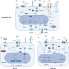
PEDF signaling in endothelial, neuronal, and RPE cells. (A) PEDF exerts its anti-angiogenic effects on proliferating endothelial cells, by binding several receptors, including LR, LRP5/6, ATP synthase, VEGFR, and PLXDC2. It generally results in endothelial cell apoptosis, downregulation of VEGF, and inhibition of angiogenesis. Apoptosis is primarily caused by PEDF binding to LR, resulting in activation of several downstream pathways and gene expression alterations, which entails activation of caspases. (B) PEDF exerts neurotrophic and neuroprotective effects in retinal neurons, mainly through binding to PEDF-R. Several downstream pathways are activated, and intracellular Ca2+ levels are decreased, which inhibits apoptosis and entails neuroprotection and survival. (C) PEDF is mainly produced in and secreted from RPE cells. However, PEDF binding to RPE cell receptors also cause downstream effects, including NPD1 production, gene expression alterations favoring apoptosis inhibition, as well as other mechanisms entailing cytoprotection. See text for details. Created with BioRender.com. PEDF, pigment epithelium-derived factor; PEDF-R, PEDF receptor; FZD. frizzled; LRP, lipoprotein receptor-related protein; LR, laminin receptor; VEGF-R, VEGF receptor; PLXDC, plexin domain containing; HIF, hypoxia-inducible factor; JNK, c-Jun N-terminal kinase; NFAT, nuclear factor of activated T-cells; Ucp-2, uncoupling protein 2; NFkB, nuclear factor kappa-light-chain-enhancer of activated B cells; PPAR-y, peroxisome proliferator-activated receptor gamma; ROS, reactive oxygen species; VEGF, vascular endothelial growth factor; c-FLIP, FLICE-like inhibitory protein; Fas-L, Fas ligand; MEK5-ERK5, mitogen-activated protein kinase 5-extracellular signal-regulated kinase 5; PMCA, plasma membrane Ca2+ ATPase; AIF, apoptosis-inducing factor; COX-2, cyclooxygenase-2; JAK, Janus kinase; STAT, signal transducers and activators of transcription; PLA2, phospholipase A2; LPL, lysophospholipids; FA, fatty acids; DHA, docosahexaenoic acid; NPD1, neuroprotection D1; PI3K, phosphoinositide 3-kinase; Akt, Akt kinase (protein kinase B); NGF, nerve growth factor; BDNF, brain-derived neurotrophic factor; GDNF, glial cell line-derived neurotrophic factor; MnSOD, manganese superoxide dismutase; Bcl, B cell lymphoma; TCF/LEF, T-cell factor/lymphoid enhancer factor.
Figure 4.
Figure 4.
Strategies for optimized delivery and efficacy of PEDF-based therapies: future perspectives for the treatment of vascular and neurodegenerative retinal disease. A major challenge for clinical application of PEDF therapeutics is efficient and durable drug delivery. PEDF therapeutics can be delivered using intraocular and periocular injections and potentially by topical application using small, derived peptides or nanocarriers. The limited ocular retention of PEDF makes direct injection clinically impractical, but advances in sustained drug delivery systems using, for example, polymer-based complexes would allow extended release with acceptable injection intervals. Truly extended therapies can be achieved using cell- and gene-based therapies. These strategies aim to achieve prolonged release of PEDF therapeutics by delivering PEDF transgenes to resident retinal cells or by modification of cells to continuously express PEDF; these cells can then be delivered to the posterior segment for engraftment or encapsulated for implantation into the vitreous. Created with BioRender.com. PEDF, pigment epithelium-derived factor.

Similar articles

References

    1. Ciulla TA, Hussain RM, Taraborelli D, Pollack JS, Williams DF.. Longer-term anti-VEGF therapy outcomes in neovascular age-related macular degeneration, diabetic macular edema, and vein occlusion-related macular edema: clinical outcomes in 130 247 eyes. Ophthalmol Retina. 2022; 6: 796–806. - PubMed
    1. Askou AL, Jakobsen TS, Corydon TJ.. Retinal gene therapy: an eye-opener of the 21st century. Gene Ther. 2021; 28: 209–216. - PubMed
    1. Dawson DW, Volpert OV, Gillis P, et al. .. Pigment epithelium-derived factor: a potent inhibitor of angiogenesis. Science. 1999; 285: 245–248. - PubMed
    1. Steele FR, Chader GJ, Johnson LV, Tombran-Tink J.. Pigment epithelium-derived factor: neurotrophic activity and identification as a member of the serine protease inhibitor gene family. Proc Natl Acad Sci USA. 1993; 90: 1526–1530. - PMC - PubMed
    1. Tombran-Tink J, Chader GG, Johnson LV.. PEDF: a pigment epithelium-derived factor with potent neuronal differentiative activity. Exp Eye Res. 1991; 53: 411–414. - PubMed

Publication types

MeSH terms

LinkOut - more resources