. 2024 Nov 24;16(12):506.
doi: 10.3390/toxins16120506.

Stable Convergent Polyneuronal Innervation and Altered Synapse Elimination in Orbicularis oculi Muscles from Patients with Blepharospasm Responding Poorly to Recurrent Botulinum Type-A Neurotoxin Injections

Affiliations

Stable Convergent Polyneuronal Innervation and Altered Synapse Elimination in Orbicularis oculi Muscles from Patients with Blepharospasm Responding Poorly to Recurrent Botulinum Type-A Neurotoxin Injections

Brigitte Girard et al. Toxins (Basel). .

Abstract

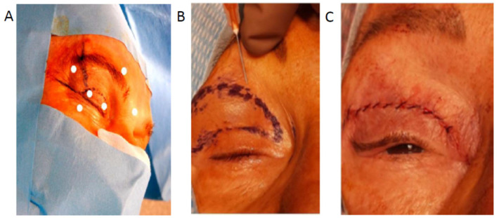
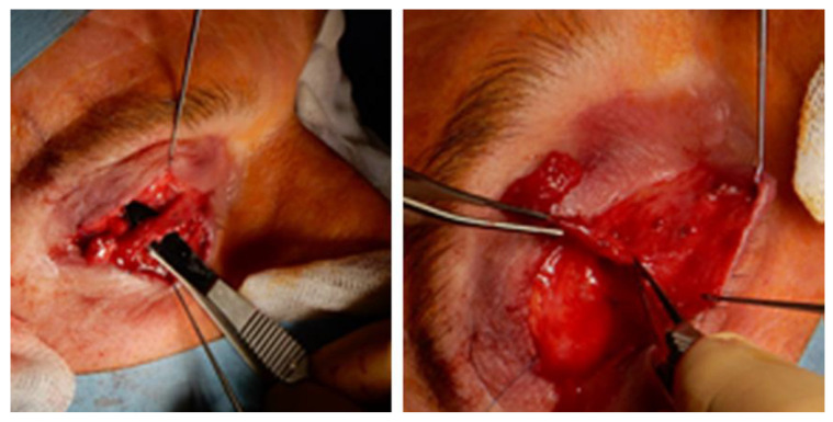
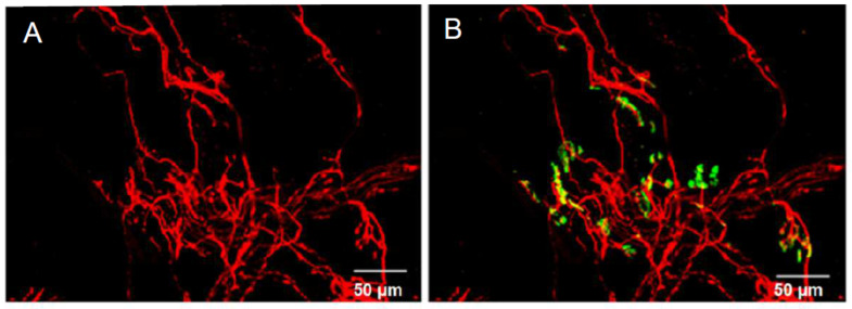
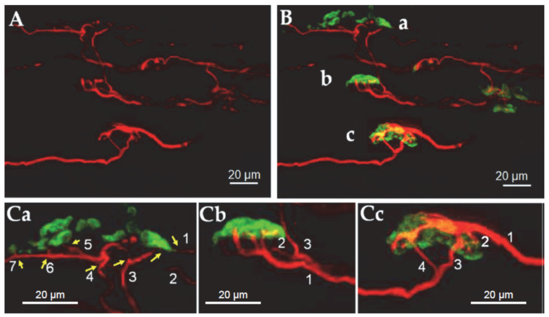
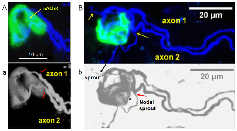
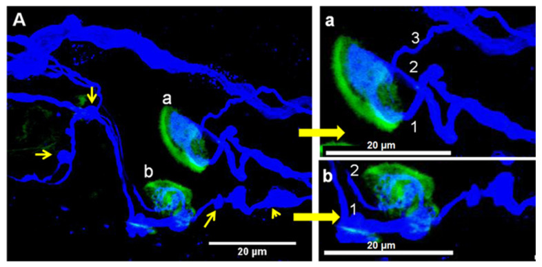
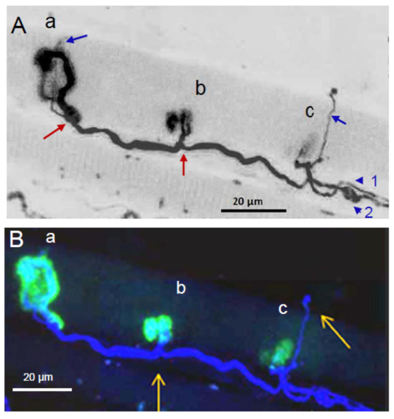
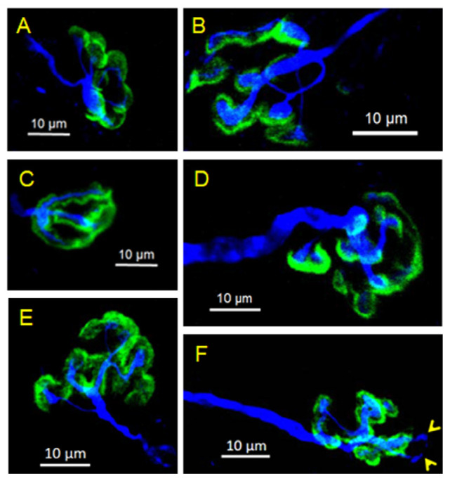
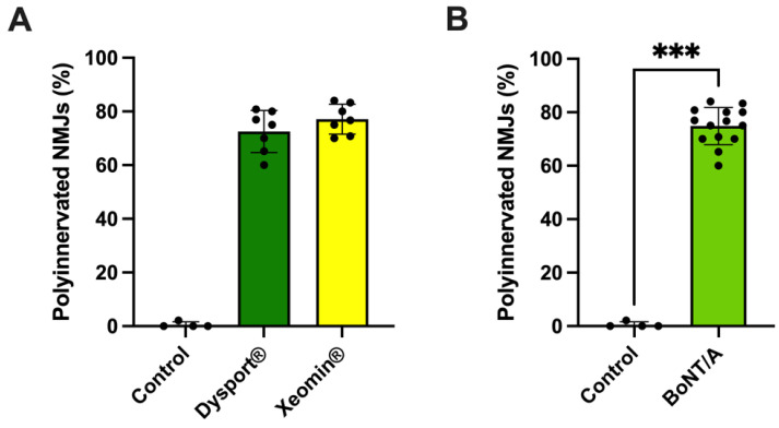
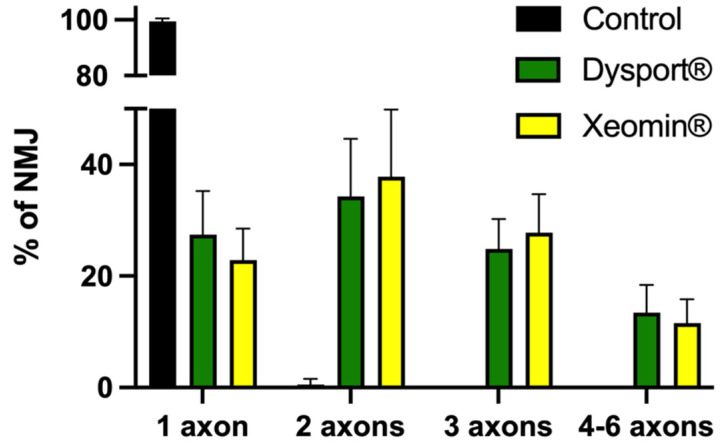
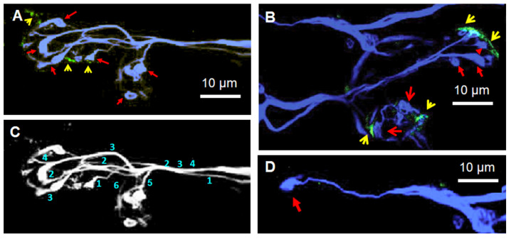
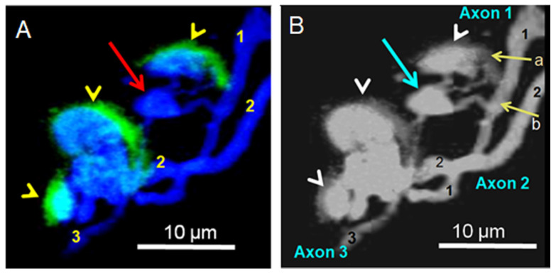
Botulinum neurotoxin type-A (BoNT/A), which blocks quantal acetylcholine (ACh) release at the neuromuscular junction (NMJ), has demonstrated its efficacy in the symptomatic treatment of blepharospasm. In 3.89% of patients treated for blepharospasm at Tenon Hospital, BoNT/A was no longer effective in relieving the patient's symptoms, and a partial upper myectomy of the Orbicularis oculi muscle was performed. We used surgical waste samples from 14 patients treated with repeated injections of either abobotulinumtoxinA (Dysport®) or incobotulinumtoxinA (Xeomin®). These muscle fragments were compared to others from 4 normal subjects, naïve of BoNT/A. The morphological study was performed blinded to the BoNT/A treatment and between treated and control samples. Neuromuscular specimens analyzed by confocal laser scanning microscopy, using fluorescent staining and immune-labeling of presynaptic proteins, revealed that the pattern of innervation (e.g., polyneuronal and convergent innervation), the muscle nicotinic ACh receptors (nAChRs), and the NMJs exhibited marked differences in BoNT/A-treated muscles (regardless of the toxin clinically used), with respect to controls. BoNT/A-treated junctions exhibited profuse polyneuronal innervation in which 2-6 axons innervated 74.84% of single muscle fibers, while 99.47% of control junctions were mono-innervated. Another new finding was the stable convergent innervation, in which several motor axons end onto the same endplate. Morphological signs of synapse elimination included the presence of retraction bulbs in axons and nerve terminals and a reduced extension of postsynaptic nAChRs. These outcomes suggest that synapse elimination is altered and raise questions on the origin and factors contributing to the plasticity changes observed and the functioning of NMJs.

Keywords: blepharospasm; botulinum type-A neurotoxin; convergent innervation; human Orbicularis oculi muscle; myectomy; nerve sprouting; neuromuscular junction; nicotinic acetylcholine receptors; polyneuronal innervation; skeletal muscle; synapse elimination.


Conflict of interest statement

The authors declare no conflict of interest. The funders had no role in the design of the study, in the collection, analyses, or interpretation of data, in the writing of the manuscript, or in the decision to publish the presented results.

Figures

Figure A1
Figure A1
(A), Outline of the injection sites of BoNT/A (white dots) in patients with essential blepharospasm at the Tenon Hospital in Paris. Note that toxin injections were done bilaterally. (B), Local anesthesia with Lidocaine injection in the drawn area. (C), Continuous cutaneous suture.
Figure A2
Figure A2
Accurate dissection of cutaneous and muscular layers to isolate and remove the preseptal Orbicularis oculi muscle above the septum.
Figure 1
Figure 1
Confocal micrographs of the innervation pattern and the endplates in a surgical-waste Orbicularis oculi mount-muscle fragment from a patient treated with BoNT/A for 1 year. In (A) are shown intramuscular axons and nerve terminals immunostained for neurofilaments (in red), and in (B) are shown both neurofilaments (in red) and nAChRs stained with α-bungarotoxin conjugated with fluorescein (in green).
Figure 2
Figure 2
The pattern of innervation and endplates imaged at high magnification with a confocal microscope from a small part of the Orbicularis oculi-mount surgical waste muscle fragment shown in Figure 1. In (A), axons and nerve terminal immunostained for neurofilaments (in red). Shown in (B) are immunostained axons and terminals innervating three different endplates labeled with fluorescent α-bungarotoxin (a, b, c, in green). Shown in (Ca,Cb,Cc) are enlarged (2.0 times) images of (B), note the polyinnervated endplates. The numbers refer to the axonal inputs (yellow arrows) to the same endplate. Note the different axonal calibers converging to single NMJs.
Figure 3
Figure 3
Examples of stable NMJs innervated by two motor axons in BoNT/A-treated Orbicularis oculi muscles. In (A,a), two myelinated axons with distinct caliber (labeled 1 and 2) converge to a unique endplate exhibiting relatively homogeneous staining with fluorescent α-bungarotoxin to label the nAChRs. Note, in (a) the heminode region (end of the myelin sheath, red arrows) of both axon 1 and axon 2. Same calibration in (A,a). (B,b) displays another complex NMJ innervated by two distinct axons. Note, in (B,b), several thin nerve terminals budding from the two axons, an ultraterminal nerve sprout (axon 1, yellow and black arrows, respectively), and a nodal sprout (axon 2, yellow and red arrows, respectively).
Figure 4
Figure 4
(A) Confocal micrograph of a representative example of convergent innervation in an Orbicularis oculi muscle fragment immunostained for neurofilaments and tubulin (blue) and stained with fluorescent α-bungarotoxin conjugated with fluorescein (yellow). The patient was treated for 1 year with BoNT/A, and the last toxin injection was performed 6 weeks before the partial myectomy. The two NMJ in (A), labeled (a,b), are shown at higher magnification in panels (a,b). The numbers 1–3 in (a) and 1, 2 in (b) refer to the axonal inputs to the respective endplates. Note in (A) the presence of oval structures connected to axons (yellow thin arrows) that may represent signs of synaptic elimination and enlarged axonal areas (yellow arrowhead).
Figure 5
Figure 5
Confocal micrographs of the innervation pattern in a surgical-waste Orbicularis oculi mount-muscle fragment, collected 6 weeks after the latest toxin injection, from a patient treated with BoNT/A for 4 years. In (A,ac) are shown two axons (blue arrowheads 1 and 2) innervating the same muscle fiber at distinct sites. In (a), note the thin nodal sprout emerging from the enlarged pre-terminal axon (red arrow) and the small terminal sprout (blue arrow). In (b) is also shown a nodal sprout (red arrow) evolving from the main nerve trunk 2. Note in (c), another axon innervating the same muscle fiber and the collateral sprout (blue arrow), budding from a nerve terminal branch. Note at the tip of the collateral sprout, there is an oval structure resembling a retraction bulb. In (B,ac), the same image as in (A), showing the fluorescent staining with α-bungarotoxin and the different extension and intensity of the labeled nAChRs (ac). The two yellow arrows show nodal and collateral sprouts on the same muscle fiber.
Figure 6
Figure 6
(AF) Confocal micrographs of the innervation pattern of Orbicularis oculi mount-muscle fragment collected from control patients that were submitted to upper blepharoplasty. These patients had neither a history of neuromuscular diseases nor blepharospasm and had never been injected with BoNT/A. Note the variability in NMJ profiles and the distinct extension of nAChR clusters labeled with fluorescent α-bungarotoxin (in green). In (F), note the occurrence of two ultraterminal short nerve sprouts (yellow arrowheads).
Figure 7
Figure 7
Percentage of polyinnervated NMJs from control patients and from patients treated with BoNT/A. (A) Percentage of polyinnervated NMJs from control patients (N = 4 donors; n = 186 NMJs examined) and from patients treated either with abobotulinumtoxinA (Dysport®, green column; N = 7, n = 160 NMJs) or with incobotulinumtoxinA (Xeomin®, yellow column; N = 7; n = 195 NMJs). Each black circle in columns represents mean data sampled from 1 patient. Data are presented as the mean ± SD. The difference between groups was statistically significant (p = 0.0029; Kruskal-Wallis test). (B) Percentage of polyinnervated NMJs from control patients (N = 4 donors; n = 186 NMJs examined) and from patients treated with abobotulinumtoxinA (Dysport®) and with incobotulinumtoxinA (Xeomin®) (Yellow Green column; N = 14, n = 355 NMJs). Data are presented as the mean ± SD. The difference between the control and BoNT/A-treated polyinnervated NMJ group was statistically significant (***) p = 0.0003); Mann-Whitney U-test.
Figure 8
Figure 8
Percentage of NMJs innervated by 1 to 6 motor axons in control patients (N = 4 donors, black column), and in patients treated with abobotulinumtoxinA (Dysport®) (N = 7; green columns), and with incobotulinumtoxinA (Xeomin®) (N = 7; yellow columns. Data is expressed as the mean ± SD. Note the significant difference between control and BoNT/A-treated NMJs innervated by 1 and 2 axons and the absence of innervation by 3 and 4–6 axons in control NMJs. No significant statistical difference was detected between the two pharmaceutical brands studied containing BoNT/A.
Figure 9
Figure 9
(AD) Signs of synaptic elimination in BoNT/A-treated O. oculi NMJs. (A,C) Presence of retraction bulbs (red arrows) with thin axonal terminals in close proximity to small clusters of nAChRs (yellow arrowheads). Note that from the six retraction bulbs, only three of them show a small nAChR cluster. (C) In the same image as in (A), the numbers indicate the axonal count in the synaptic region. (B) Two polyinnervated NMJs from another BoNT/A-treated patient showed nAChR clusters (yellow arrowheads) and some retraction bulbs, some of which are devoid of nAChR clusters (red arrows). (D) Unique retraction bulb (red arrow) attached to an atrophied thin axonal extension emerging from a polyaxonal intramuscular nerve trunk.
Figure 10
Figure 10
(A,B) Single NMJ innervated by three distinct axons (numbered 1–3) showing a retraction bulb (red and turquoise arrows in (A,B), respectively) located in between innervated nAChR clusters stained with fluorescent α-bungarotoxin (yellow arrowheads). Note in (B) that the retraction bulb is connected to at least two nodal axonal branches emerging from the axon 1 (indicated by the yellow arrows labeled a and b, respectively).

Similar articles

Cited by

References

    1. Pirazzini M., Montecucco C., Rossetto O. Toxicology and pharmacology of botulinum and tetanus neurotoxins: An update. Arch. Toxicol. 2022;96:1521–1539. doi: 10.1007/s00204-022-03271-9. - DOI - PMC - PubMed
    1. Cai S., Kumar R., Singh B.R. Clostridial Neurotoxins: Structure, Function and Implications to Other Bacterial Toxins. Microorganisms. 2021;9:2206. doi: 10.3390/microorganisms9112206. - DOI - PMC - PubMed
    1. Poulain B., Popoff M.R. Why Are Botulinum Neurotoxin-Producing Bacteria So Diverse and Botulinum Neurotoxins So Toxic? Toxins. 2019;11:34. doi: 10.3390/toxins11010034. - DOI - PMC - PubMed
    1. Hill K.K., Xie G., Foley B.T., Smith T.J. Genetic diversity within the botulinum neurotoxin-producing bacteria and their neurotoxins. Pt AToxicon. 2015;107:2–8. doi: 10.1016/j.toxicon.2015.09.011. - DOI - PubMed
    1. Johnson E.A. Clostridial toxins as therapeutic agents: Benefits of nature’s most toxic proteins. Annu. Rev. Microbiol. 1999;53:551–575. doi: 10.1146/annurev.micro.53.1.551. - DOI - PubMed

LinkOut - more resources