Review
. 2024 Dec 20;10(12):2100-2127.
doi: 10.3390/tomography10120149.

Pediatric Neuroimaging of Multiple Sclerosis and Neuroinflammatory Diseases

Affiliations
Review

Pediatric Neuroimaging of Multiple Sclerosis and Neuroinflammatory Diseases

Chloe Dunseath et al. Tomography. .

Abstract

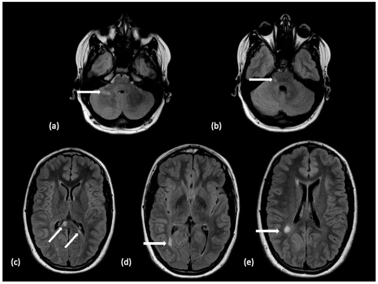
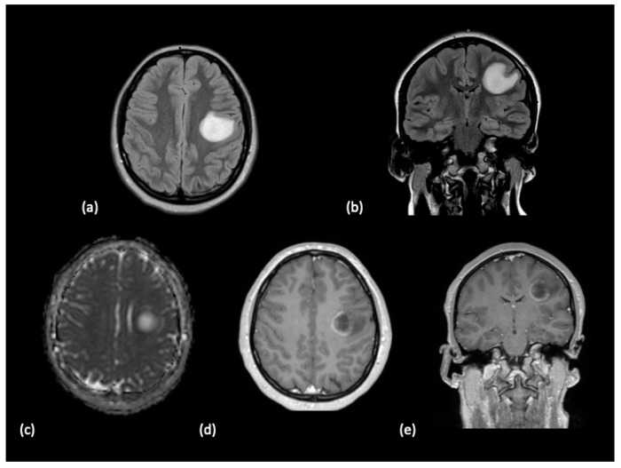
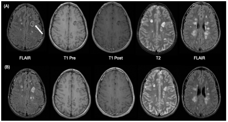
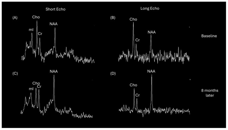
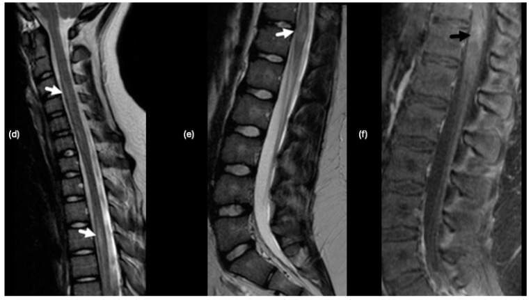
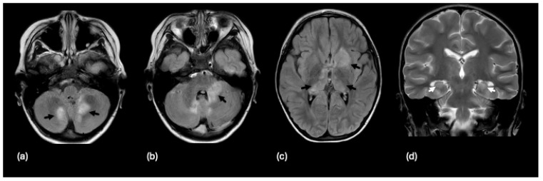
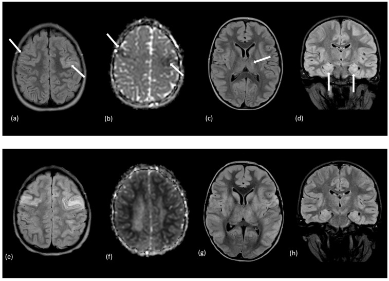
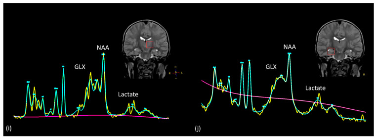
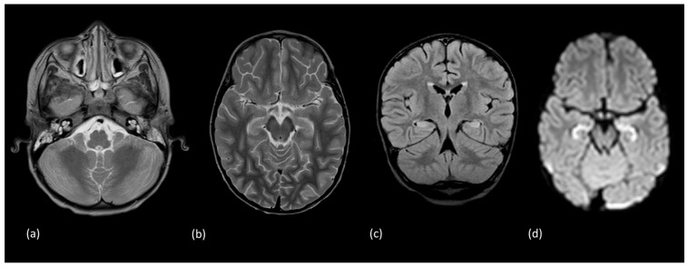
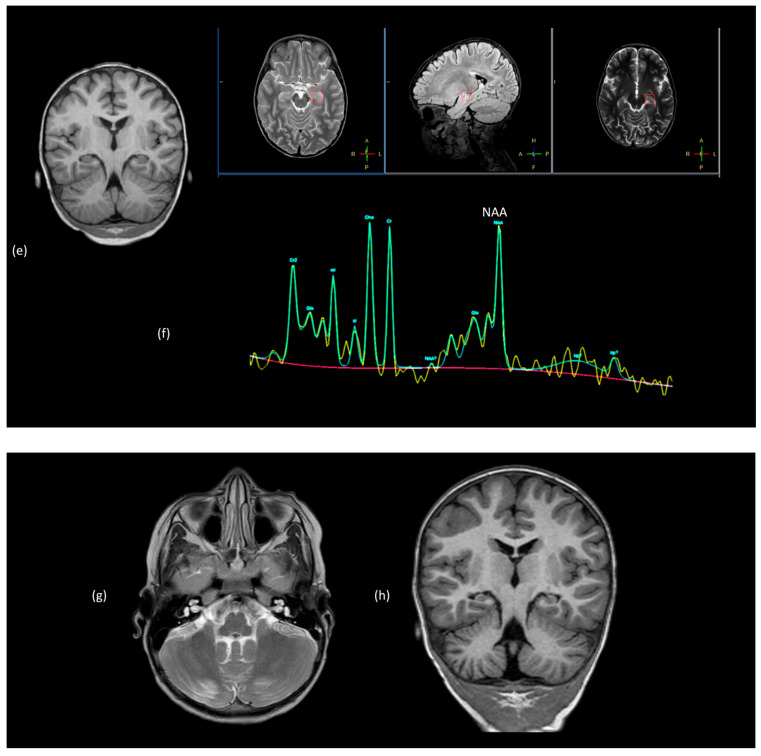
Using a pediatric-focused lens, this review article briefly summarizes the presentation of several demyelinating and neuroinflammatory diseases using conventional magnetic resonance imaging (MRI) sequences, such as T1-weighted with and without an exogenous gadolinium-based contrast agent, T2-weighted, and fluid-attenuated inversion recovery (FLAIR). These conventional sequences exploit the intrinsic properties of tissue to provide a distinct signal contrast that is useful for evaluating disease features and monitoring treatment responses in patients by characterizing lesion involvement in the central nervous system and tracking temporal features with blood-brain barrier disruption. Illustrative examples are presented for pediatric-onset multiple sclerosis and neuroinflammatory diseases. This work also highlights findings from advanced MRI techniques, often infrequently employed due to the challenges involved in acquisition, post-processing, and interpretation, and identifies the need for future studies to extract the unique information, such as alterations in neurochemistry, disruptions of structural organization, or atypical functional connectivity, that may be relevant for the diagnosis and management of disease.

Keywords: acute demyelinating encephalomyelitis; demyelination; magnetic resonance imaging; magnetic resonance spectroscopy; multiple sclerosis; myelin oligodendrocyte glycoprotein antibody disease; neuroinflammatory.


Conflict of interest statement

The authors declare no conflicts of interest.

Figures

Figure 1
Figure 1
A 14-year-old male with a 3-year history of multiple demyelinating, non-enhancing brain lesions. A follow-up MRI demonstrated an increase in both size and number of lesions with a distribution within the brain characteristic of pediatric-onset multiple sclerosis. Non-enhancing lesions on an axial FLAIR sequence without diffusion restriction are noted with white arrows within the (a) right hemisphere cerebellar white matter, (b) dorsal right pons, (c) splenium of the corpus callosum, (d) bordering the right occipital horn, and (e) right hemispheric parietal white matter. The features supported the diagnosis of pediatric-onset multiple sclerosis.
Figure 2
Figure 2
A 15-year-old female with a history of bilateral optic neuritis presented with stroke-like symptoms including facial droop and slurred speech. (a) Axial and (b) coronal FLAIR sequences demonstrated a large hyperintense lesion located in the left middle frontal lobes, precentral gyrus and the frontal operculum. (c) The lesion exhibits mild diffusion restriction at the periphery. Partial peripheral enhancement along the medial margin of the lesion was noted on the (d) axial and (e) coronal post-contrast T1-weighted images. The patient history and imaging features over time supported a diagnosis of tumefactive multiple sclerosis.
Figure 3
Figure 3
A 14-year-old male who presented with a history of headaches and vague numbness in his right upper limb. Row (A) demonstrates initial baseline imaging and Row (B) shows follow-up imaging 8 months later. From left to right in each row are FLAIR, T1-weighted pre-contrast, T1-weighted post-contrast, T2-weighted axial images at the level of a concentric ring lesion (white arrow) in the left frontal lobe and a FLAIR image at a superior level illustrating periventricular lesions. This appearance is consistent with Balo’s concentric sclerosis. Comparison of corresponding images in rows A and B, show many lesions were smaller at follow-up imaging than on the prior baseline study. None of the patient’s lesions significantly enhanced after contrast administration at either timepoint on T1-weighted imaging.
Figure 4
Figure 4
Short echo (35 milliseconds) and long echo (288 milliseconds) single voxel proton MRS acquired in the left frontal lobe Balo’s lesion of a 14-year-old male at initial baseline and follow-up examination 8 months later for the patient described in Figure 3. Short echo MRS in (A) demonstrates elevated choline-to-creatine and myo-inositol-to-creatine ratios compared with ratios observed in (C). Long echo MRS in (B) also reflects the choline elevation. The spectra normalize as shown in (C,D) with lower levels of choline and higher N-acetyl-aspartate.
Figure 5
Figure 5
A 7-year-old male presented with an acute history of fever, ataxia, and dysarthria. Brain MRI demonstrated multiple hyperintense signals on T2/FLAIR sequences that displayed faint hypointense signals on T1-weighted sequences, without diffusion restriction, abnormal contrast agent enhancement or mass effect. Lesions are noted with white arrows. (a) Lesions within the pons and cerebral peduncle are hyperintense on an axial T2-weighted image. (b) Sagittal T1-weighted and (c) axial FLAIR feature a lesion in the left cerebellar hemisphere. (d,e) Axial FLAIR images demonstrate bilateral thalamic and left centrum semiovale lesions, respectively. (f) An additional ill-defined, T2-mildly hyperintense lesion was noted at the T9–10 of the spinal cord. This lesion was without abnormal enhancement on T1-weighted imaging. These imaging features supported the diagnosis of acute demyelinating encephalomyelitis (ADEM).
Figure 6
Figure 6
A 13-year-old female presenting with bilateral papilledema and imaging findings of optic neuritis. Coronal T2-weighted image (a) with fat saturation demonstrates bilateral optic nerve edema and swelling (white arrows). Coronal (b) and axial (c) fat-saturated, post-contrast T1-weighted images demonstrate abnormal enhancement (white arrows). The authors formally obtained permission to use clinically generated medical images stored within the Cincinnati Children’s Hospital Medical Center’s PACS for this figure on 6 December 2024.
Figure 7
Figure 7
A 10-year-old male presenting with left lower limb weakness, pain, and hyperreflexia. (a) Axial FLAIR image demonstrates a non-enhancing, small, focal hyperintense signal abnormality along the posterior inferior medulla (white arrow). On coronal FLAIR (b,c), there is asymmetric edema in the right optic nerve (white arrow) and subtle swelling and signal abnormality of the right optic chiasm (white arrowhead). (d) 2-year-follow-up MR shows resolution of medullary signal abnormality (black arrow) on axial FLAIR.
Figure 8
Figure 8
A 10-year-old female presenting with right monocular vision loss. (a) Coronal T2 orbital image demonstrates abnormal edema and swelling of the intraorbital right optic nerve (white arrow), as well as abnormal enhancement (black arrows) of the (b) prechiasmatic (b,c) intraorbital right optic nerve. Six weeks later, the patient presents with new voiding and extremity weakness. On (d,e) sagittal T2-weighted images, there are ill-defined cord signal abnormalities (white arrows) with associated subtle enhancement (black arrow) of the distal cord on (f) sagittal T1-weighted post contrast fat saturated spine MR image.
Figure 8
Figure 8
A 10-year-old female presenting with right monocular vision loss. (a) Coronal T2 orbital image demonstrates abnormal edema and swelling of the intraorbital right optic nerve (white arrow), as well as abnormal enhancement (black arrows) of the (b) prechiasmatic (b,c) intraorbital right optic nerve. Six weeks later, the patient presents with new voiding and extremity weakness. On (d,e) sagittal T2-weighted images, there are ill-defined cord signal abnormalities (white arrows) with associated subtle enhancement (black arrow) of the distal cord on (f) sagittal T1-weighted post contrast fat saturated spine MR image.
Figure 9
Figure 9
A 4-year-old male with recent viral encephalitis presenting with new gait ataxia, finger tingling, and headache and positive MOG antibody. Axial FLAIR images demonstrate multifocal, non-enhancing lesions (black arrows) in the (a) cerebellum, (b) left brachium pontis, and (c) deep gray matter structures, including the left caudate, anterior left putamen and globus pallidus, and the right greater than left thalami. Additional bilateral hippocampal swelling and signal abnormalities (white arrows) are shown on (d) coronal T2-weighted images.
Figure 10
Figure 10
A 6-year-old male presented with new onset seizures, facial droop, and a family history of transient ischemic attacks. The initial MRI with (a) axial FLAIR image and (b) diffusion ADC map demonstrated multifocal areas of abnormal signal bilaterally (white arrows) involving the superior frontal gyri. Axial FLAIR imaging also revealed (c) a left thalamic lesion, and (d) hippocampal formations showing increased signal intensity and enlargement without diffusion restriction. Select FLAIR images from a follow-up study performed a week later showed progression throughout (e) frontal cortices, (g) thalamus and insular cortices, and (h) hippocampi. The (f) ADC map demonstrated restriction throughout subcortical white matter. Additional lesions emerged in the parietal lobes, pons, cerebellar white matter, thalami, and basal ganglia. MRS (i,j) with voxel placement illustrated on coronal T2 images featured elevated signal for lactate (1.3 ppm) and glutamate/glutamine (GLX, 2.1–2.5 ppm) accompanied by reduced N-acetyl aspartate (NAA) (2.0 ppm) levels for the left thalamic and right hippocampal regions sampled. These features supported the diagnosis of autoimmune encephalitis.
Figure 10
Figure 10
A 6-year-old male presented with new onset seizures, facial droop, and a family history of transient ischemic attacks. The initial MRI with (a) axial FLAIR image and (b) diffusion ADC map demonstrated multifocal areas of abnormal signal bilaterally (white arrows) involving the superior frontal gyri. Axial FLAIR imaging also revealed (c) a left thalamic lesion, and (d) hippocampal formations showing increased signal intensity and enlargement without diffusion restriction. Select FLAIR images from a follow-up study performed a week later showed progression throughout (e) frontal cortices, (g) thalamus and insular cortices, and (h) hippocampi. The (f) ADC map demonstrated restriction throughout subcortical white matter. Additional lesions emerged in the parietal lobes, pons, cerebellar white matter, thalami, and basal ganglia. MRS (i,j) with voxel placement illustrated on coronal T2 images featured elevated signal for lactate (1.3 ppm) and glutamate/glutamine (GLX, 2.1–2.5 ppm) accompanied by reduced N-acetyl aspartate (NAA) (2.0 ppm) levels for the left thalamic and right hippocampal regions sampled. These features supported the diagnosis of autoimmune encephalitis.
Figure 11
Figure 11
A 3-year-old male presented with acute onset seizure. Initial MRI demonstrated increased signal intensity and diffusion restriction involving both hippocampal formations with subtle signal abnormality involving the adjacent amygdala. (a) Axial T2-weighted imaging at the level of the cerebellum. (b) Axial T2-weighted, (c) coronal FLAIR, and (d) axial diffusion-weighted imaging reveal hyperintense signal within the hippocampus. (e) Initial coronal T1-weighted imaging. (f) Short echo magnetic resonance spectroscopy of the left hippocampus demonstrated reduced N-acetyl aspartate (resonance located at 2.0 ppm). In a follow-up study performed 6 months later demonstrated resolution of the noted hippocampal signal findings. However, asymmetric volume loss of the cerebellum with prominent cerebellar sulci ((g) axial T2-weighted) and right hippocampal formation ((h) coronal T1-weighted) imaging. These features were consistent with a diagnosis of febrile-infection-related epilepsy syndrome (FIRES).
Figure 11
Figure 11
A 3-year-old male presented with acute onset seizure. Initial MRI demonstrated increased signal intensity and diffusion restriction involving both hippocampal formations with subtle signal abnormality involving the adjacent amygdala. (a) Axial T2-weighted imaging at the level of the cerebellum. (b) Axial T2-weighted, (c) coronal FLAIR, and (d) axial diffusion-weighted imaging reveal hyperintense signal within the hippocampus. (e) Initial coronal T1-weighted imaging. (f) Short echo magnetic resonance spectroscopy of the left hippocampus demonstrated reduced N-acetyl aspartate (resonance located at 2.0 ppm). In a follow-up study performed 6 months later demonstrated resolution of the noted hippocampal signal findings. However, asymmetric volume loss of the cerebellum with prominent cerebellar sulci ((g) axial T2-weighted) and right hippocampal formation ((h) coronal T1-weighted) imaging. These features were consistent with a diagnosis of febrile-infection-related epilepsy syndrome (FIRES).

Similar articles

References

    1. Alroughani R., Boyko A. Pediatric multiple sclerosis: A review. BMC Neurol. 2018;18:27. doi: 10.1186/s12883-018-1026-3. - DOI - PMC - PubMed
    1. Krupp L.B., Vieira M.C., Toledano H., Peneva D., Druyts E., Wu P., Boulos F.C. A Review of Available Treatments, Clinical Evidence, and Guidelines for Diagnosis and Treatment of Pediatric Multiple Sclerosis in the United States. J. Child. Neurol. 2019;34:612–620. doi: 10.1177/0883073819855592. - DOI - PubMed
    1. Boiko A., Vorobeychik G., Paty D., Devonshire V., Sadovnick D., University of British Columbia M.S.C.N. Early onset multiple sclerosis: A longitudinal study. Neurology. 2002;59:1006–1010. doi: 10.1212/WNL.59.7.1006. - DOI - PubMed
    1. Patel Y., Bhise V., Krupp L. Pediatric multiple sclerosis. Ann. Indian. Acad. Neurol. 2009;12:238–245. doi: 10.4103/0972-2327.58281. - DOI - PMC - PubMed
    1. Prajjwal P., Marsool M.D.M., Natarajan B., Inban P., Gadam S., Sowndarya D., John J., Abbas R., Vaja H., Marsool M.B., et al. Juvenile multiple sclerosis: Addressing epidemiology, diagnosis, therapeutic, and prognostic updates along with cognitive dysfunction and quality of life. Ann. Med. Surg. 2023;85:4433–4441. doi: 10.1097/MS9.0000000000000930. - DOI - PMC - PubMed

MeSH terms

LinkOut - more resources