Automated Measurement of Effective Radiation Dose by 18F-Fluorodeoxyglucose Positron Emission Tomography/Computed Tomography
- PMID: 39728913
- PMCID: PMC11679132
- DOI: 10.3390/tomography10120151
Automated Measurement of Effective Radiation Dose by 18F-Fluorodeoxyglucose Positron Emission Tomography/Computed Tomography
Abstract
Background/objectives: Calculating the radiation dose from CT in 18F-PET/CT examinations poses a significant challenge. The objective of this study is to develop a deep learning-based automated program that standardizes the measurement of radiation doses.
Methods: The torso CT was segmented into six distinct regions using TotalSegmentator. An automated program was employed to extract the necessary information and calculate the effective dose (ED) of PET/CT. The accuracy of our automated program was verified by comparing the EDs calculated by the program with those determined by a nuclear medicine physician (n = 30). Additionally, we compared the EDs obtained from an older PET/CT scanner with those from a newer PET/CT scanner (n = 42).
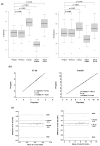
Results: The CT ED calculated by the automated program was not significantly different from that calculated by the nuclear medicine physician (3.67 ± 0.61 mSv and 3.62 ± 0.60 mSv, respectively, p = 0.7623). Similarly, the total ED showed no significant difference between the two calculation methods (8.10 ± 1.40 mSv and 8.05 ± 1.39 mSv, respectively, p = 0.8957). A very strong correlation was observed in both the CT ED and total ED between the two measurements (r2 = 0.9981 and 0.9996, respectively). The automated program showed excellent repeatability and reproducibility. When comparing the older and newer PET/CT scanners, the PET ED was significantly lower in the newer scanner than in the older scanner (4.39 ± 0.91 mSv and 6.00 ± 1.17 mSv, respectively, p < 0.0001). Consequently, the total ED was significantly lower in the newer scanner than in the older scanner (8.22 ± 1.53 mSv and 9.65 ± 1.34 mSv, respectively, p < 0.0001).
Conclusions: We successfully developed an automated program for calculating the ED of torso 18F-PET/CT. By integrating a deep learning model, the program effectively eliminated inter-operator variability.
Keywords: 18F-FDG; computed tomography; deep learning; effective dose; positron emission tomography.
Conflict of interest statement
The authors declare no conflicts of interest.
Figures


References
-
- UNEP . What Is Radiation? What Does Radiation Do to Us? Where Does Radiation Come from? UNSCEAR; Vienna, Austria: 2016. Radiation Effects and Sources; pp. 1–10, 13–17, 27–37. UNED, ed.
-
- Richardson D.B., Leuraud K., Laurier D., Gillies M., Haylock R., Kelly-Reif K., Bertke S., Daniels R.D., Thierry-Chef I., Moissonnier M., et al. Cancer mortality after low dose exposure to ionising radiation in workers in France, the United Kingdom, and the United States (INWORKS): Cohort study. BMJ. 2023;382:e074520. doi: 10.1136/bmj-2022-074520. - DOI - PMC - PubMed
Publication types
MeSH terms
Substances
Grants and funding
LinkOut - more resources
Full Text Sources

