Case Reports
. 2024 Dec 7;11(12):632.
doi: 10.3390/vetsci11120632.

Intestinal Osteosarcoma with Liver Metastasis in a Dog with a History of Recurrent Cotton-Based Toy Fragment Ingestion

Affiliations
Case Reports

Intestinal Osteosarcoma with Liver Metastasis in a Dog with a History of Recurrent Cotton-Based Toy Fragment Ingestion

Andrada Negoescu et al. Vet Sci. .

Abstract

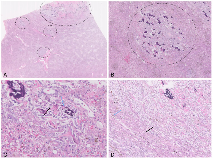
Canine extraskeletal osteosarcomas are mesenchymal, osteoid producing tumors that can arise in soft tissues without initial involvement of the bones. An 8-year-old intact male Beagle dog presented with anorexia, abdominal pain, intermittent vomiting and melena. The patient had a history of recurrent ingestion of cotton based-toy fragments, but no prior surgical procedures involving the abdominal cavity. During the exploratory laparotomy, a mass was identified in the jejunal wall. Surgical resection was performed, and tissue samples were collected for pathological examination. Histologically, the mass was diagnosed as osteoblastic osteosarcoma with fragments of cotton fiber material. The neoplastic cells were immunolabeled for vimentin and BMP-2, further supporting the morphological diagnosis. Seven months after the surgery, metastatic nodules were identified in the liver. The dog died ten months after intestinal mass resection. This case represents the first documented instance of metastatic intestinal osteosarcoma potentially caused by ingestion of cotton fiber material.

Keywords: cotton fibers; dog; extraskeletal osteosarcoma; immunohistochemistry; intestine.


Conflict of interest statement

The authors declare no conflicts of interest.

Figures

Figure 1
Figure 1
Ultrasound and macroscopical features of the canine intestinal osteosarcoma. The mass is characterized by a hypoechoic external region and an anechoic center (measured area 2); in the top right corner there is a normal intestinal loop (measured area 1); (A). Grossly, the intestinal wall is severely distended by a poorly delimited, infiltrative and dense mass (white arrow) (B).
Figure 2
Figure 2
Photomicrographs of the canine intestinal osteosarcoma. The jejunum is transmurally infiltrated and replaced by a neoplastic mass containing cotton fiber fragments (the inset) (A). The neoplastic cells are polygonal to spindle, arranged in interwoven streams and bundles, associated with numerous islands of homogenous acidophilic extracellular matrix/osteoid (black arrow) partially mineralized (blue arrow) (B), confirmed by MT stain (black arrow) (C). The fragments of foreign material exhibit birefringence under polarized light (arrow) (D). Immunohistochemically, the neoplastic cells exhibit a strong cytoplasmatic expression for vimentin (E), and moderate cytoplasmatic expression for BMP 2 (blue arrows) (F).
Figure 3
Figure 3
Photomicrographs of the metastatic osteosarcoma in the liver. The hepatic parenchyma is multifocally expanded and replaced by variably sized neoplastic nodules (delimited areas) (A,B). The tumor cells (black arrow) are intermingled with small islands of partially mineralized osteoid (blue arrow) (C). The adjacent parenchyma is compressed, contains small lipogranulomas (blue arrow) and the hepatocytes show vacuolar degeneration/lipidosis (black arrow) (D).

Similar articles

Cited by

  • Focus on Tumours in Pet Animals.
    van der Weyden L. van der Weyden L. Vet Sci. 2025 Mar 12;12(3):266. doi: 10.3390/vetsci12030266. Vet Sci. 2025. PMID: 40266980 Free PMC article.

References

    1. Slovak J.E., Kieves N.R., Haynes J. Extraskeletal Osteosarcoma Induced by a Foreign Body Granuloma. J. Am. Anim. Hosp. Assoc. 2015;51:315–319. doi: 10.5326/JAAHA-MS-6224. - DOI - PubMed
    1. Silver K.I., Patkar S., Mazcko C., Berger E.P., Beck J.A., LeBlanc A.K. Patterns of Metastatic Progression and Association with Clinical Outcomes in Canine Osteosarcoma: A Necropsy Study of 83 Dogs. Vet. Comp. Oncol. 2023;21:646–655. doi: 10.1111/vco.12927. - DOI - PMC - PubMed
    1. Egenvall A., Nødtvedt A., von Euler H. Bone Tumors in a Population of 400,000 Insured Swedish Dogs Up to 10 Years of Age: Incidence and Survival. Can. J. Vet. Res. 2007;71:292–299. - PMC - PubMed
    1. Langenbach A., Anderson M.A., Dambach D.M., Sorenmo K.U., Shofer F.D. Extraskeletal Osteosarcomas in Dogs: A Retrospective Study of 169 Cases (1986–1996) J. Am. Anim. Hosp. Assoc. 1998;34:113–120. doi: 10.5326/15473317-34-2-113. - DOI - PubMed
    1. McMahon M., Mathie T., Stingle N., Romansik E., Vail D., London C. Adjuvant Carboplatin and Gemcitabine Combination Chemotherapy Postamputation in Canine Appendicular Osteosarcoma. J. Vet. Intern. Med. 2011;25:511–517. doi: 10.1111/j.1939-1676.2011.0697.x. - DOI - PMC - PubMed

Publication types

LinkOut - more resources