. 2025 Feb;14(2):391-411.
doi: 10.1007/s40123-024-01077-9. Epub 2024 Dec 27.

Detection of Retinal and Choriocapillaris Microvascular Changes in Retinal Vein Occlusion and Fellow Eyes by Optical Coherence Tomography Angiography: A Systematic Review and Meta-Analysis

Affiliations

Detection of Retinal and Choriocapillaris Microvascular Changes in Retinal Vein Occlusion and Fellow Eyes by Optical Coherence Tomography Angiography: A Systematic Review and Meta-Analysis

Linxin Wei et al. Ophthalmol Ther. 2025 Feb.

Abstract

Introduction: This study aims to summarize the retinal and choroidal microvascular features detected by optical coherence tomography angiography (OCTA) in the affected and fellow eyes of patients with retinal vein occlusion (RVO).

Methods: A comprehensive search of the PubMed, Embase, and Ovid databases was conducted to identify studies comparing OCTA metrics among RVO, RVO-fellow, and control eyes. Outcomes of interest included parameters related to foveal avascular zone (FAZ) and fovea- and optic nerve head (ONH)-centered perfusion measurements of superficial capillary plexus (SCP), deep capillary plexus (DCP), and choriocapillaris layer. Pooled results were presented as mean differences or standardized mean differences with 95% confidence intervals.

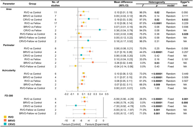
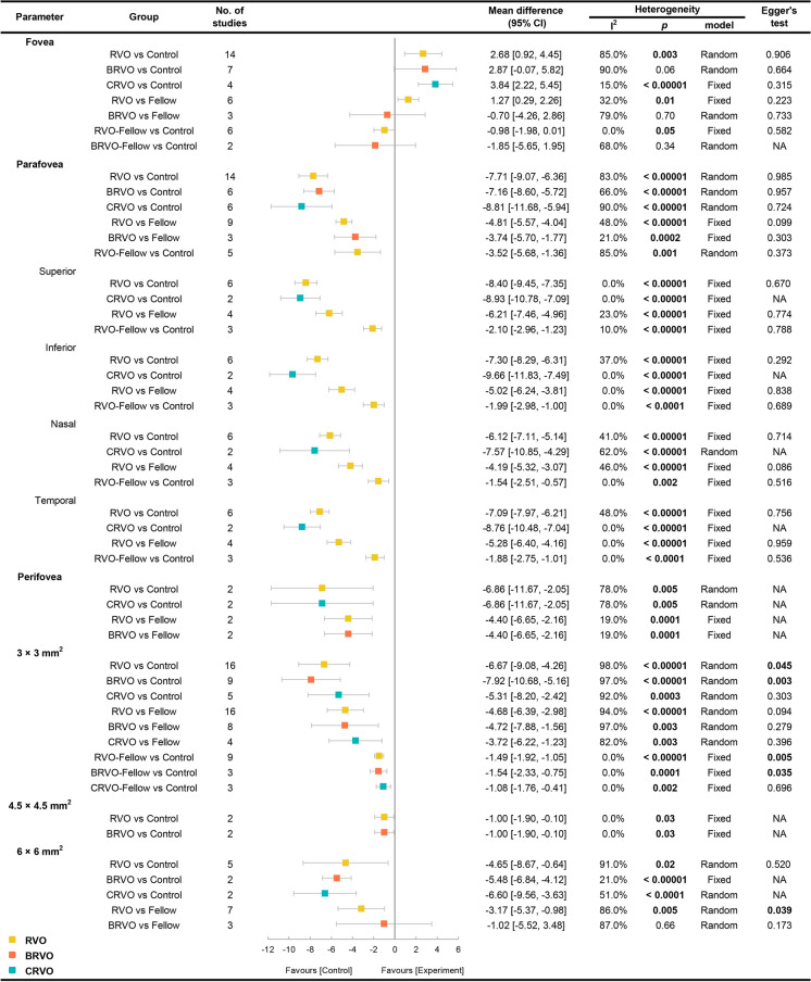
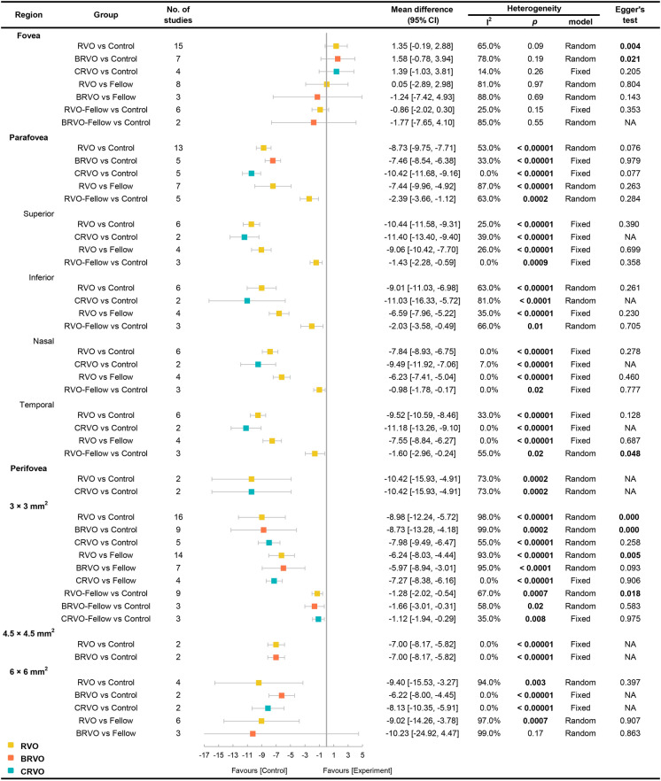
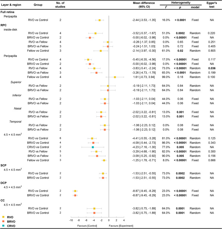
Results: Fifty-three studies, comprising 2119 RVO, 1393 fellow, and 1178 control eyes, were included in the quantitative meta-analysis. RVO eyes exhibited larger FAZ areas, increased FAZ acircularity, and reduced macular retinal and choriocapillaris perfusion compared to RVO-fellow and control eyes (P < 0.05). RVO eyes also demonstrated significantly lower perfusion density (PD) in the inside-disk and peripapillary regions of the radial peripapillary capillary layer (RPC), as well as lower retinal and choriocapillaris PD in the 4.5 × 4.5 mm2 field of view (FOV) of ONH-centered scans (P < 0.05). RVO-fellow eyes showed decreased SCP-PD and DCP-PD in the parafoveal region and the 3 × 3 mm2 FOV, reduced inside-disk and 4.5 × 4.5 mm2 FOV RPC-PD (P < 0.05), and a diminished choriocapillaris flow area in the 3 × 3 mm2 FOV (P < 0.05).

Conclusions: Both RVO-affected and RVO-fellow eyes exhibited retinal and choriocapillaris microvascular impairment around the fovea and ONH. OCTA represents a promising tool for comprehensively assessing vascular alterations in RVO and providing evidence of fellow eye involvement.

Keywords: Choroid; Meta-analysis; Optical coherence tomography angiography; Retina; Retinal vein occlusion.


Conflict of interest statement

Declarations. Conflict of Interest: Linxin Wei, Qing Zhao, and Youxin Chen declare that they have no competing interests. Ethical Approval: This article is based on previously conducted studies and does not contain any new studies with human participants or animals performed by any of the authors.

Figures

Fig. 1
Fig. 1
Flowchart of the study selection
Fig. 2
Fig. 2
Group comparisons of FAZ metrics in OCTA measurements. OCTA parameters including FAZ area, perimeter, acircularity and FD-300 are compared among RVO, fellow, control eyes, whose results are displayed by mean difference and 95% CI. The publication bias of each pooled result is shown by Egger’s linear regression test result. The bold font indicates that the result is significant. When RVO eyes are compared to fellow or control eyes, RVO eyes are called “Experiment” and the other two groups are called “Control”; when fellow eyes are compared to control eyes, fellow eyes are called “Experiment” and controls are called “Control”. BRVO retinal vein occlusion, CI confidence interval, CRVO central retinal vein occlusion, DCP deep capillary plexus, FAZ foveal avascular zone, FD-300 PD within a 300-μm-wide annulus surrounding FAZ, OCTA optical coherence tomography angiography, RVO retinal vein occlusion, SCP superficial capillary plexus
Fig. 3
Fig. 3
Group comparisons of SCP-PD in OCTA measurements. Measured subregions for SCP-PD include the fovea, parafovea (further divided as superior, inferior, nasal, and temporal areas), perifovea, 3 × 3 mm2, 4.5 × 4.5 mm2, and 6 × 6 mm2, whose results are displayed by mean difference and 95% CI. The publication bias of each pooled result is shown by Egger’s linear regression test result. The bold font indicates that the result is significant. When RVO eyes are compared to fellow or control eyes, RVO eyes are called “Experiment” and the other two groups are called “Control”; when fellow eyes are compared to control eyes, fellow eyes are called “Experiment” and controls are called “Control”. BRVO retinal vein occlusion, CI confidence interval, CRVO central retinal vein occlusion, OCTA optical coherence tomography angiography, PD perfusion density, RVO retinal vein occlusion, SCP superficial capillary plexus
Fig. 4
Fig. 4
Group comparisons of DCP-PD in OCTA measurements. Measured subregions for DCP-PD include the fovea, parafovea (further divided as superior, inferior, nasal, and temporal areas), perifovea, 3 × 3 mm2, 4.5 × 4.5 mm2, and 6 × 6 mm2, whose results are displayed by mean difference and 95% CI. The publication bias of each pooled result is shown by Egger’s linear regression test result. The bold font indicates that the result is significant. When RVO eyes are compared to fellow or control eyes, RVO eyes are called “Experiment” and the other two groups are called “Control”; when fellow eyes are compared to control eyes, fellow eyes are called “Experiment” and controls are called “Control”. BRVO retinal vein occlusion, CI confidence interval, CRVO central retinal vein occlusion, DCP deep capillary plexus, OCTA optical coherence tomography angiography, PD perfusion density, RVO retinal vein occlusion
Fig. 5
Fig. 5
Group comparisons of ONH-PD in OCTA measurements. Measured layers and subregions for ONH-PD include the peripapilla for the full-retina layer, the inside-disk, peripapilla (further divided as superior, inferior, nasal, and temporal areas), and 4.5 × 4.5 mm2 for the RPC layer, and 4.5 × 4.5 mm2 for the SCP, DCP, and CC layers. The comparison results are displayed by mean difference and 95% CI. The publication bias of each pooled result is shown by Egger’s linear regression test result. The bold font indicates that the result is significant. When RVO eyes are compared to fellow or control eyes, RVO eyes are called “Experiment” and the other two groups are called “Control”; when fellow eyes are compared to control eyes, fellow eyes are called “Experiment” and controls are called “Control”. BRVO retinal vein occlusion, CC choriocapillaris, CI confidence interval, CRVO central retinal vein occlusion, DCP deep capillary plexus, OCTA optical coherence tomography angiography, PD perfusion density, RPC radial peripapillary capillary, RVO retinal vein occlusion, SCP superficial capillary plexus

Similar articles

References

    1. Song P, Xu Y, Zha M, Zhang Y, Rudan I. Global epidemiology of retinal vein occlusion: a systematic review and meta-analysis of prevalence, incidence, and risk factors. J Glob Health. 2019;9(1): 010427. - PMC - PubMed
    1. Marcinkowska A, Cisiecki S, Rozalski M. Platelet and thrombophilia-related risk factors of retinal vein occlusion. J Clin Med. 2021;10(14):3080. - PMC - PubMed
    1. Yin S, Cui Y, Jiao W, Zhao B. Potential prognostic indicators for patients with retinal vein occlusion. Front Med (Lausanne). 2022;9:839082. - PMC - PubMed
    1. Özçalışkan Ş, Özcan Y, Artunay Ö. Macular optical coherence tomography angiography parameters in central retinal vein occlusion and differentiation of ischemic and non-ischemic types. Haseki Tip Bulteni. 2021;59(1):68–73.
    1. Chen L, Yuan M, Sun L, Wang Y, Chen Y. Evaluation of microvascular network with optical coherence tomography angiography (OCTA) in branch retinal vein occlusion (BRVO). BMC Ophthalmol. 2020;20(1):154. - PMC - PubMed

LinkOut - more resources