. 2024 Dec 27;19(12):e0316199.
doi: 10.1371/journal.pone.0316199. eCollection 2024.

Construction of reusable fluorescent assembled 3D-printed hydrogen-based models to simulate minimally invasive resection of complex liver cancer

Affiliations

Construction of reusable fluorescent assembled 3D-printed hydrogen-based models to simulate minimally invasive resection of complex liver cancer

Wenli Cao et al. PLoS One. .

Abstract

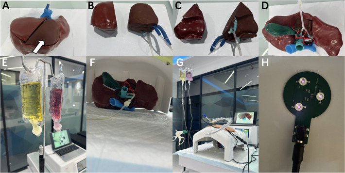
Complex liver cancer is often difficult to expose or dissect, and the surgery is often challenging. 3D-printed models may realistically present 3D anatomical structure, which has certain value in planning and training of liver surgery. However, the existing 3D-printed models are all monolithic models, which are difficult to reuse and limited in clinical application. It is also rare to carry fluorescence to accurately present tumor lesions. Here we report reusable fluorescent assembled 3D-printed models to mimic minimally invasive resection of complex liver cancer. Based on the models, multiple copies of liver lesion structure assembled accessories can be printed for the same patient or different patients, ensuring the quantity and quality of simulated surgical training, and greatly reducing the cost of simulated surgical training. The addition of fluorescence is helpful in accurately presenting tumor lesions. The reusable fluorescent assembled 3D-printed models may mimic minimally invasive resection of complex liver cancer, demonstrating potential value in simulated surgery.


Conflict of interest statement

None reported

Figures

Fig 1
Fig 1. Workflow of simulated surgery using reusable fluorescent assembled 3D-printed hydrogen-based models.
Fig 2
Fig 2. Design and creation of 3D liver models.
A: The electronic design of the middle liver assembled block (marked by the blue arrow) with a liver tumor located at segment 4a (marked by the yellow arrow). Additionally, an immobilized liver block has been designed (marked by the purple circle). B: The electronic design of the right liver assembled block (marked by the blue triangle) incorporates a smaller, movable assembled block (marked by the green triangle). Within that smaller block, a liver tumor is located at segment 8 (marked by the yellow triangle). Additionally, an immobilized liver block has been designed (marked by the purple circle). C: Creation of real 3D silicone-based models for the liver immobilization block (marked by the red circle), with two movable segments (marked by the red triangle and arrow) of the posterior superior region of the liver, allowing for future replacement with hydrogel models. D: Addition of auxiliary structures such as gallbladder, hepatic artery to make the model more complete.
Fig 3
Fig 3. Preparation of models and simulated surgery.
A,D: The hydrogel-based assembled block (marked by the white arrow) is put into the reserved groove in the silicone-based immobilized block. B,C: The silicone-based assembled block, and the hydrogel-based assembled block with the hepatic vein and portal vein branches connecting to small tubes. E: The infusion bags carrying different stained saline to simulate the blood. F: The models were placed in an abdominal cavity. G: The hepatic vein and portal vein branches in the hydrogel-based 3D models of liver assembled block are connected with infusion bags containing simulated blood to simulate blood flow. And the models were placed in an abdominal cavity simulator with a camera connected to the display screen to simulate the surgical process. H: Homemade infrared light for detection of fluorescence.
Fig 4
Fig 4. The models were used to simulate surgery for resection of liver tumor located at segment 4a.
A-C: Electronic design of 3D image of the liver with liver tumor lesion at segment 4a. D,E: Models prepared well for simulated laparoscopic surgery. F,G: The hydrogel-based block (marked by the white arrow) was treated by electrocoagulation. H,I: Laparoscopic suture of the hepatic vein branch. J,K: Laparoscopic liver resection was almost completed gradually to reveal the liver tumor. L: Laparoscopic suture of the portal vein branch. M: The wound after mass removal. N,O: Intraoperative fluorescence navigation to show the tumor boundary. P,Q: After the tumor is removed, fluorescence irradiation can stain the entire tumor and its boundaries.
Fig 5
Fig 5. The models were used to simulate surgery for resection of liver tumor located at segment 8.
A-D: Electronic design of 3D image of the liver with liver tumor lesion at segment 8. E: The whole silicone-based assembled block, and the hydrogel-based assembled block (marked by the white triangle) is embedded in the silicone-based assembled block. F,G: Models prepared well for simulated laparoscopic surgery. H: The hydrogel-based block(marked by the white triangle) was treated by electrocoagulation. I,J: Laparoscopic suture of the portal vein branch. K: Laparoscopic liver resection was almost completed gradually to reveal the liver tumor. L: Laparoscopic suture of the hepatic vein branch. M: The mass was completely removed. N,O: Intraoperative fluorescence navigation to show the tumor boundary. P,Q: After the tumor is removed, fluorescence irradiation can stain the entire tumor and its boundaries.

References

    1. Sheng W., Yuan C., Wu L., et al.. (2022). Clinical application of a three-dimensional reconstruction technique for complex liver cancer resection. Surg Endosc.36(5), 3246–3253. doi: 10.1007/s00464-021-08636-2 - DOI - PubMed
    1. Shi W., Wang J., Gao J., et al.. (2024).Utilization of 3D printing technology in hepatopancreatobiliary surgery. J Zhejiang Univ Sci B.25(2), 123–134. - PMC - PubMed
    1. Yang T., Lin S., Xie Q., et al.. (2019). Impact of 3D printing technology on the comprehension of surgical liver anatomy. Surg Endosc.33(2), 411–417. doi: 10.1007/s00464-018-6308-8 - DOI - PubMed
    1. Lupulescu C., & Sun Z. (2019). A Systematic Review of the Clinical Value and Applications of Three-Dimensional Printing in Renal Surgery. J Clin Med. 8(7), 990. doi: 10.3390/jcm8070990 - DOI - PMC - PubMed
    1. Bastawrous S., Wake N., Levin D., et al.. (2018). Principles of three-dimensional printing and clinical applications within the abdomen and pelvis. Abdom Radiol (NY). 43(10), 2809–2822. doi: 10.1007/s00261-018-1554-8 - DOI - PubMed

MeSH terms