

Low grade appendiceal mucinous neoplasm mimicking malignant ovarian tumor: A case report
- PMID: 39729895
- PMCID: PMC11741048
- DOI: 10.1016/j.ijscr.2024.110767
Low grade appendiceal mucinous neoplasm mimicking malignant ovarian tumor: A case report
Abstract
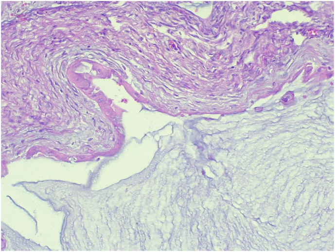
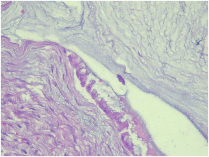
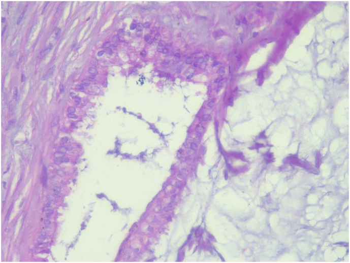
Introduction: Mucinous appendiceal neoplasms are unique tumors in which >50 % of the tumor volume is composed of extracellular mucin. They may present as an unruptured mucin-filled appendix or, more commonly, with peritoneal metastases after rupture or transmural invasion of the primary tumor. This case report describes a case of presumed ovarian malignancy with final pathologic diagnosis of low grade appendiceal mucinous neoplasm. Due to its rarity, we decided to report it.
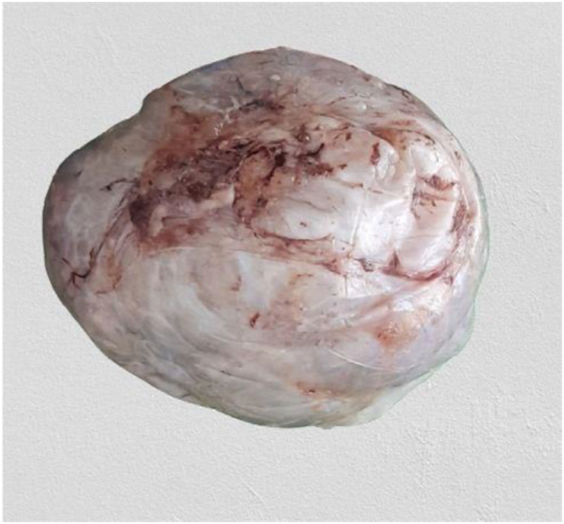
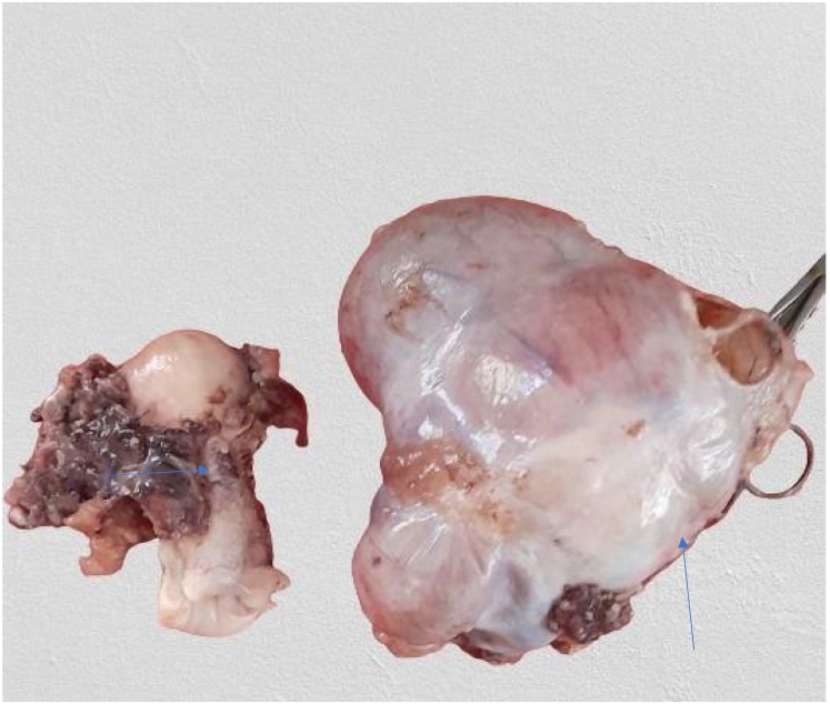
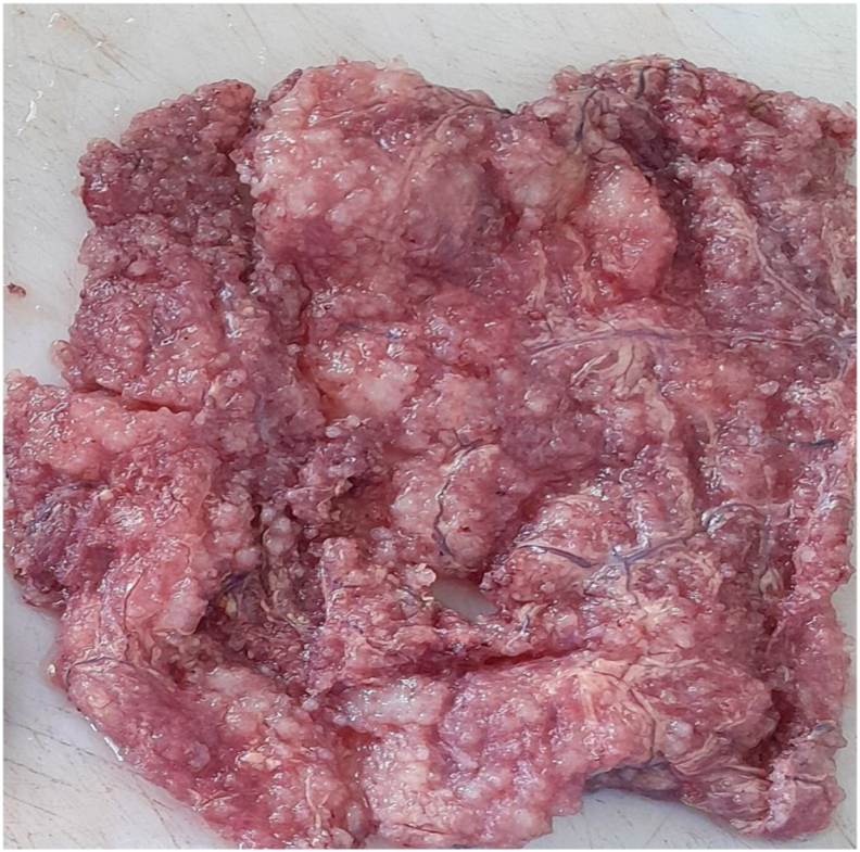
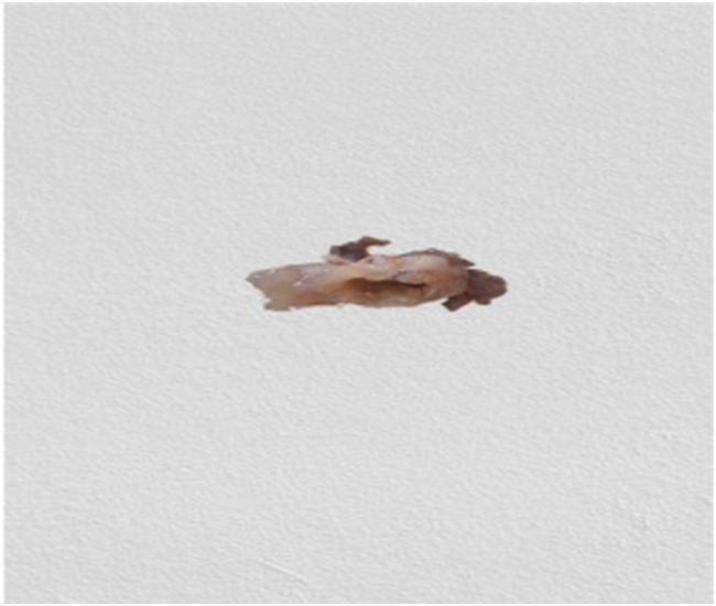
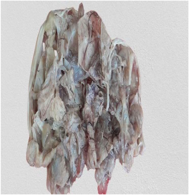
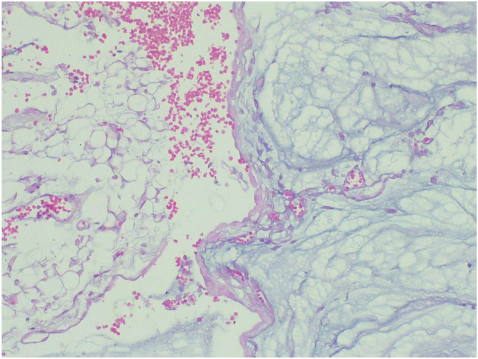
Case presentation: A 37-year-old patient presented with a compliant of abdominal swelling. Abdominopelvic ultrasound was done and showed huge right and left complex cystic ovarian masses having thick septa. For this, she underwent total abdominal hysterectomy, omentectomy and bilateral salpingo-oophorectomy. On laparotomy there was also incidental finding of ruptured mucin-filled appendix for which appendectomy was done. Histopathology examinations from all resected specimens revealed the diagnosed LAMN. Two weeks after surgical resection, she was started on FOLFOX chemotherapy regimen.
Discussion: Incidence of low grade appendiceal mucinous neoplasm is increasing. In addition to the increasing incidence, lack of early detection and impeded access to optimal multi-disciplinary treatment may worsen survival outcomes. Developing quality diagnostic services in the proper health context is crucial for early diagnosis and successful therapy of LAMN patients, and applying a resource-sensitive approach to prioritize essential treatments based on effectiveness and cost-effectiveness is key to overcoming barriers in low- and middle-income countries.
Conclusion: A recognition of mucinous material and abnormal appearing appendix should prompt the surgeon to consider performing an appendectomy to obtain primary pathologic diagnosis.
Keywords: Case report; Low grade appendiceal mucinous neoplasm; Low- and middle-income countries; Ovarian cancer; Pseudomyxoma peritonei.
Copyright © 2024 The Authors. Published by Elsevier Ltd.. All rights reserved.
Conflict of interest statement
Declaration of competing interest N/A
Figures
Similar articles
-
Low grade appendiceal mucinous neoplasm metastatic to the ovary: A case report and intraoperative assessment guide.Int J Surg Case Rep. 2023 Aug;109:108563. doi: 10.1016/j.ijscr.2023.108563. Epub 2023 Jul 26. Int J Surg Case Rep. 2023. PMID: 37524024 Free PMC article.
-
Low-grade appendiceal mucinous neoplasm associated with Urothelial carcinoma: A rare case report from Syria.Ann Med Surg (Lond). 2022 Mar 29;76:103525. doi: 10.1016/j.amsu.2022.103525. eCollection 2022 Apr. Ann Med Surg (Lond). 2022. PMID: 35495395 Free PMC article.
-
Low-grade appendiceal mucinous neoplasm encountered during risk-reducing salpingo-oophorectomy: A case of laparoscopic surgery.J Obstet Gynaecol Res. 2023 Dec;49(12):2975-2978. doi: 10.1111/jog.15802. Epub 2023 Sep 28. J Obstet Gynaecol Res. 2023. PMID: 37771102
-
[Incidental finding of appendiceal mucinous neoplasms].Chirurgie (Heidelb). 2023 Oct;94(10):832-839. doi: 10.1007/s00104-023-01910-0. Epub 2023 Jun 28. Chirurgie (Heidelb). 2023. PMID: 37378666 Review. German.
-
Pseudomyxoma peritonei and selected other aspects of the spread of appendiceal neoplasms.Semin Diagn Pathol. 2004 May;21(2):134-50. doi: 10.1053/j.semdp.2004.12.002. Semin Diagn Pathol. 2004. PMID: 15807473 Review.
References
-
- Umetsu S.E., Kakar S. Staging of appendiceal mucinous neoplasms: challenges and recent updates. Hum. Pathol. 2023 Feb 1;132:65–76. - PubMed
-
- Reiter Shelby, et al. Progression to pseudomyxoma peritonei in patients with low grade appendiceal mucinous neoplasms discovered at time of appendectomy. Am. J. Surg. 2022;223(6):1183–1186. - PubMed
-
- Misdraji J. Mucinous epithelial neoplasms of the appendix and pseudomyxoma peritonei. Mod. Pathol. 2015 Jan 1;28:S67–S79. - PubMed
Publication types
LinkOut - more resources
Full Text Sources
Medical
