. 2024 Dec 27;9(1):371.
doi: 10.1038/s41392-024-02077-8.

Itaconate facilitates viral infection via alkylating GDI2 and retaining Rab GTPase on the membrane

Affiliations

Itaconate facilitates viral infection via alkylating GDI2 and retaining Rab GTPase on the membrane

Shulei Yin et al. Signal Transduct Target Ther. .

Abstract

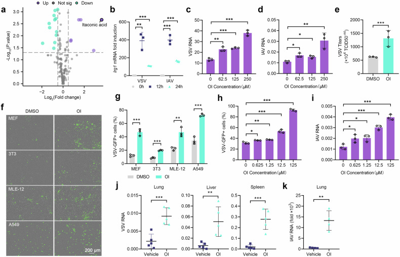
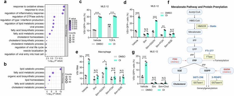
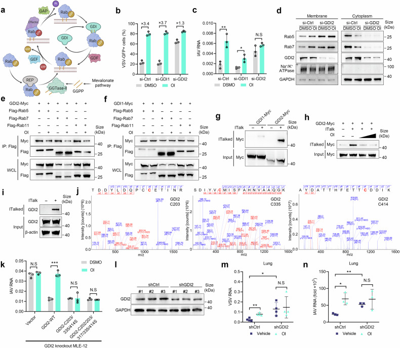
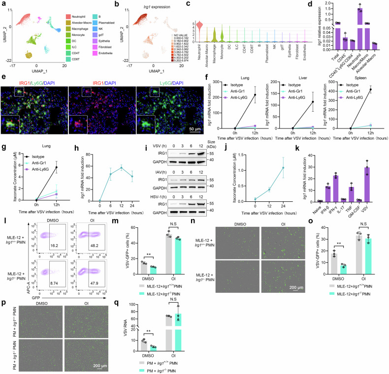
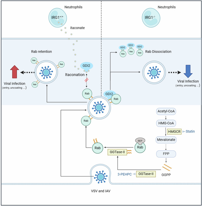
Metabolic reprogramming of host cells plays critical roles during viral infection. Itaconate, a metabolite produced from cis-aconitate in the tricarboxylic acid cycle (TCA) by immune responsive gene 1 (IRG1), is involved in regulating innate immune response and pathogen infection. However, its involvement in viral infection and underlying mechanisms remain incompletely understood. Here, we demonstrate that the IRG1-itaconate axis facilitates the infections of VSV and IAV in macrophages and epithelial cells via Rab GTPases redistribution. Mechanistically, itaconate promotes the retention of Rab GTPases on the membrane via directly alkylating Rab GDP dissociation inhibitor beta (GDI2), the latter of which extracts Rab GTPases from the membrane to the cytoplasm. Multiple alkylated residues by itaconate, including cysteines 203, 335, and 414 on GDI2, were found to be important during viral infection. Additionally, this effect of itaconate needs an adequate distribution of Rab GTPases on the membrane, which relies on Rab geranylgeranyl transferase (GGTase-II)-mediated geranylgeranylation of Rab GTPases. The single-cell RNA sequencing data revealed high expression of IRG1 primarily in neutrophils during viral infection. Co-cultured and in vivo animal experiments demonstrated that itaconate produced by neutrophils plays a dominant role in promoting viral infection. Overall, our study reveals that neutrophils-derived itaconate facilitates viral infection via redistribution of Rab GTPases, suggesting potential targets for antiviral therapy.


Conflict of interest statement

Competing interests: The authors declare no competing interests.

Figures

Fig. 1
Fig. 1
Inducible IRG1-itaconate axis facilitates viral infection. a Metabolites in the lungs from mice i.p. infected with VSV or not (n = 6). b Irg1 mRNA expression in the lungs of mice intranasally (i.n.) infected with VSV and IAV (n = 3). c, d Viral RNA in PMs pretreated with different concentrations of OI, infected with VSV (c) or IAV (d) (n = 3). e Viral titers in the supernatant of PMs pretreated with OI (250 μM) or DMSO (n = 3). f, g VSV-GFP infection in MEF, 3T3, MLE-12 and A549 cells pretreated with OI (125 μM) or DMSO, detected by fluorescence (f) and flow cytometry (g) (n = 3). h, i VSV-GFP (h) and IAV (i) infection in MLE-12 cells pretreated with different concentrations of OI. j, k Mice were pretreated with OI, subsequently infected with VSV (i.p.) and IAV (i.n.), and viral RNA loads were detected (n = 5 or 4). Data are mean ± SD or representative of 3 independent experiments with similar results. *p < 0.05, **p < 0.01, ***p < 0.001 by an unpaired, two-tailed t-test
Fig. 2
Fig. 2
IRG1 facilitates VSV and IAV infection independent of IFN-I pathway. a The most significantly altered metabolic genes upon VSV infection were identified by RNA-seq (n = 2). b Irg1 mRNA expression in PMs infected with VSV, IAV or HSV-1 for 8 h (n = 3). c Immunoblot of IRG1 in PMs infected with VSV or IAV. d Itaconate in the supernatants of PMs infected with VSV for 12 h (n = 3). e, f Viral RNA in Irg1+/+ and Irg1-/- BMDMs infected with VSV (e) or IAV (f) (n = 3). g ELISA analysis of IFN-β in the supernatants of Irg1+/+ and Irg1-/- BMDMs infected with VSV for 24 h (n = 3). h Isg15 and Mx1 mRNA expression in Irg1+/+ and Irg1-/- BMDMs infected with VSV (n = 3). i Immunoblot of IRG1, RIG-I, MAVS, TBK1, phosphorylated TBK1 (p-TBK1), IRF3, phosphorylated IRF3 (p-IRF3), STAT1, phosphorylated STAT1 (p-STAT1) in Irg1+/+ and Irg1-/- BMDMs infected with VSV. j, k Ifnb1 (j) and Isg15 (k) mRNA expression in Irg1+/+ and Irg1-/- BMDMs infected with IAV (n = 3). l Ifnb1 mRNA expression in Irg1+/+ and Irg1-/- BMDMs transfected with poly I:C (3 ug/ml) for 8 h (n = 3). m Ifnb1 and Isg15 mRNA expression in Irg1+/+ and Irg1-/- BMDMs infected with UV-VSV (n = 3). n VSV RNA in Irf3-/- PMs carrying Irg1 or control siRNA (n = 3). o HSV-1 RNA in Irg1+/+ and Irg1-/- BMDMs (n = 3). p ELISA analysis of IFN-β in the supernatants of Irg1+/+ and Irg1-/- BMDMs infected with HSV-1 for 24 h (n = 3). q VSV RNA in Irg1+/+ and Irg1-/- BMDMs pretreated with OI (250 μM) or DMSO (n = 3). r The effect of OI on VSV-GFP infection rate in Irf3-/- PMs carrying Irg1 or control siRNA (n = 3). Data are mean ± SD or representative of 3 independent experiments with similar results. *p < 0.05, **p < 0.01, ***p < 0.001, N.S, not significant by an unpaired, two-tailed t-test
Fig. 3
Fig. 3
GGPP is indispensable for facilitating viral infection induced by OI. a, b GO Pathway enrichment analysis of differentially expressed genes from the RNA-seq data of Irg1+/+ and Irg1-/- BMDMs (a), and PMs treated with OI (250 μM) or DMSO (b). c The effect of OI on VSV-GFP infection rate in MLE-12 cells pretreated with TOFA (30 μM) or vehicle (n = 3). d, e The effect of OI on VSV infection in MLE-12 cells (d) and PMs (e) pretreated with simvastatin (Sim, 10 μM) plus GGOH (15 μM), FOH (15 μM), SQE (15 μM) or not (n = 3). f Schematic of mevalonate pathway and protein prenylation. g The effect of OI on VSV-GFP infection rate in MLE-12 cells pretreated with simvastatin (Sim, 10 μM) with or without a cell permeable cholesterol (Chol, 5 ug/ml) (n = 3). Data are mean ± SD. *p < 0.05, **p < 0.01, ***p < 0.001, N.S, not significant by an unpaired, two-tailed t-test
Fig. 4
Fig. 4
OI induces the redistribution of Rab GTPases to enhance viral infection. a Immunoblot of GGTase-II substrate proteins in the membrane, cytoplasm and total cell fractions of MLE-12 cells pretreated with OI (125 μM) or DMSO. b Confocal of Rab5 and EEA1 co-localization (n = 20), Rab7 and LAMP1 co-localization (n = 23), in MLE-12 cells pretreated with OI or DMSO. The icon on the right displays the co-localization rate. c The effect of OI on Rab5 and Rab7 distribution in the membrane and cytoplasm in MLE-12 cells treated with simvastatin (Sim, 10 μM), 3-PEHPC (1.5 mM) or vehicle as indicated. d, e VSV-GFP (d) and IAV (e) infection in MLE-12 cells pretreated with OI or DMSO plus FTI-277 (10 μM), GGTI-298 (10 μM), 3-PEHPC (1.5 mM) or vehicle for 12 h (n = 3). f VSV RNA in PMs and MLE-12 cells pretreated with OI or DMSO 1 h post infection (n = 3). g VSV RNA in Irg1+/+ and Irg1-/- BMDMs infected with VSV for 1 h (n = 3). h, i VSV RNA in MLE-12 cells pretreated with OI or DMSO (h), and in Irg1+/+ and Irg1-/- BMDMs (i) infected with VSV for 1 h at 4 °C (n = 3). j, k VSV RNA in MEF cells pretreated with OI or DMSO (j), and in Irg1+/+ and Irg1-/- MEF cells (k) transfected with VSV-RNA (n = 3). l, m The effect of OI on VSV-GFP (l) and IAV (m) infection in MLE-12 cells silenced of Rab5, Rab7, or Rab11 (n = 3). Data are mean ± SD or representative of 3 independent experiments with similar results. *p < 0.05, **p < 0.01, ***p < 0.001, N.S, not significant by an unpaired, two-tailed t-test
Fig. 5
Fig. 5
OI inhibits the extraction of Rabs from the membrane via alkylation of GDI2. a Schematic of Rab GTPases cycle. b, c The effect of OI on VSV-GFP (b) and IAV (c) infection in MLE-12 cells silenced of GDI1 or GDI2 (n = 3). d Immunoblot of Rab5 and Rab7 in the membrane and cytoplasm of MLE-12 cells carrying GDI2 or control siRNA, and treated with OI (125 μM) or DMSO. e, f The effect of OI on the interaction of GDI2 (e) and GDI1 (f) with the Rabs in HEK293T cells transfected with GDI2-Myc or GDI1-Myc together with Flag-Rab5, Flag-Rab7 or Flag-Rab11. g Immunoblot of ITalk-modified (ITalked) GDI1 and GDI2 in HEK293T cells transfected with GDI1-Myc or GDI2-Myc. h Immunoblot of ITalked GDI2 in HEK293T cells treated with different concentrations of OI (0, 62.5 or 125 μM). i ITalked endogenous GDI2 proteins in MLE-12 cells treated with ITalk or DMSO. j LC-MS/MS analysis of itaconation on GDI2 in HEK293T cells transfected with GDI2-Flag, treated with OI for 12 h. k The effect of OI on IAV infection in GDI2-deficient MLE-12 cells transfected with wildtype or mutated GDI2 (n = 3). l Immunoblot of GDI2 in the lungs from mice intratracheally administered with adeno-associated virus containing GDI2 shRNA (shGDI2) or scrambled shRNA (shCtrl) (n = 3 per group). m VSV RNA in the lungs of mice upon GDI2 knockdown and control mice (n = 4). n IAV RNA in the lungs of mice upon GDI2 knockdown and control mice (n = 3). Data are mean ± SD or representative of 3 independent experiments with similar results. *p < 0.05, **p < 0.01, ***p < 0.001, N.S, not significant by an unpaired, two-tailed t-test
Fig. 6
Fig. 6
Viral infection induces robust IRG1-itaconate axis activation in neutrophils. a, b UMAP clusters (a) and Irg1 expression (b) of scRNA-seq data obtained from the lung of mouse i.p. infected with VSV for 12 h. c Violin plots showing Irg1 expression in each cluster. d Irg1 mRNA expression in different cells sorted from the VSV infected lungs (n = 3). e Immunofluorescence analysis of IRG1 and Ly6G expression in VSV infected lungs. f, g Irg1 mRNA expression in the lungs, livers and spleens (f), and itaconate accumulation in the lung (g) of neutrophil-depleted and control mice (n = 3). h Irg1 mRNA expression in neutrophils infected with VSV (n = 3). i Immunoblot of IRG1 in neutrophils infected with VSV, IAV or HSV-1. j Itaconate in the supernatant of neutrophils infected with VSV (n = 3). k Irg1 mRNA expression in neutrophils stimulated with IFN-β (100 ng/ml), IFN-γ (100 ng/ml), IL-17 (100 ng/ml), TNF (100 ng/ml), GM-CSF (100 ng/ml) or VSV for 8 h (n = 3). lo VSV-GFP infection rate in MLE-12 cells co-cultured with Irg1+/+ or Irg1-/- neutrophils (ratio = 1:1), pretreated with OI or DMSO (l, m) (n = 3). A transwell system was used in (n, o) (n = 3). p, q VSV-GFP infection in PMs co-cultured with Irg1+/+ or Irg1-/- neutrophils as in (l), was revealed through fluorescence (p), qRT-PCR (q) (n = 3). Data are mean ± SD or representative of 3 independent experiments with similar results. **p < 0.01, N.S, not significant by an unpaired, two-tailed t-test
Fig. 7
Fig. 7
Neutrophils-derived itaconate facilitates VSV and IAV infection. a The model of chimeric mice experiments. b Itaconate in the lungs of chimeric mice infected with VSV or IAV (n = 2 or 5). c VSV RNA in the lungs, spleens and livers of chimeric mice (n = 5). d ELISA analysis of IFN-β, IL-6 and TNF-α in the serum of mice from (c) (n = 5). e H&E of lung sections of mice from (b). f IAV RNA in the lungs of chimeric mice (n = 5). g Ifnb1, Il6 and Tnf mRNA expression in the lungs of mice from (f) (n = 5). h The model of neutrophils deleting experiments. i VSV RNA in the lungs, spleens and livers of neutrophils-deleted and control mice (n = 5). j ELISA analysis of IFN-β, IL-6 and TNF-α in the serum of mice from (i) (n = 5). k H&E of lung sections of mice from (i). l IAV RNA in the lungs of neutrophils-deleted and control mice (n = 4 or 5). m Ifnb1 and Il6 mRNA expression in the lungs of mice from (l) (n = 4 or 5). Data are mean ± SD or representative of 3 independent experiments with similar results. *p < 0.05, **p < 0.01, ***p < 0.001, N.S, not significant by an unpaired, two-tailed t-test
Fig. 8
Fig. 8
Neutrophils-derived itaconate facilitates viral infection through inducing Rab GTPases redistribution. VSV and IAV infections induce a robust activation of the IRG1-itaconate axis in neutrophils. Subsequently, the secreted itaconate enters macrophages and epithelial cells, where it alkylates GDI2 and impedes its extraction of Rab GTPases from the membrane. As a result, there is an increased retention of Rabs on the membrane, ultimately facilitating Rabs-dependent viral infection

References

    1. Girdhar, K. et al. Viruses and metabolism: the effects of viral infections and viral insulins on host metabolism. Annu Rev. Virol.8, 373–391 (2021). - PMC - PubMed
    1. Moreno-Altamirano, M. M. B., Kolstoe, S. E. & Sánchez-García, F. J. Virus control of cell metabolism for replication and evasion of host immune responses. Front. Cell Infect. Microbiol.9, 95 (2019). - PMC - PubMed
    1. Thaker, S. K., Ch’ng, J. & Christofk, H. R. Viral hijacking of cellular metabolism. BMC Biol.17, 59 (2019). - PMC - PubMed
    1. Palmer, ClovisS. Innate metabolic responses against viral infections. Nat. Metab.4, 1245–1259 (2022). - PubMed
    1. Michelucci, A. et al. Immune-responsive gene 1 protein links metabolism to immunity by catalyzing itaconic acid production. Proc. Natl Acad. Sci. USA110, 7820–7825 (2013). - PMC - PubMed

Publication types

MeSH terms