. 2024 Dec 28;14(1):30786.
doi: 10.1038/s41598-024-80837-w.

A multiparametric screen uncovers FDA-approved small molecules that potentiate the nuclear mechano-dysfunctions in ATR-defective cells

Affiliations

A multiparametric screen uncovers FDA-approved small molecules that potentiate the nuclear mechano-dysfunctions in ATR-defective cells

Maria Rosaria Cera et al. Sci Rep. .

Abstract

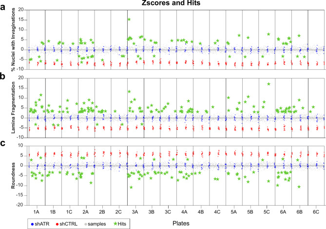
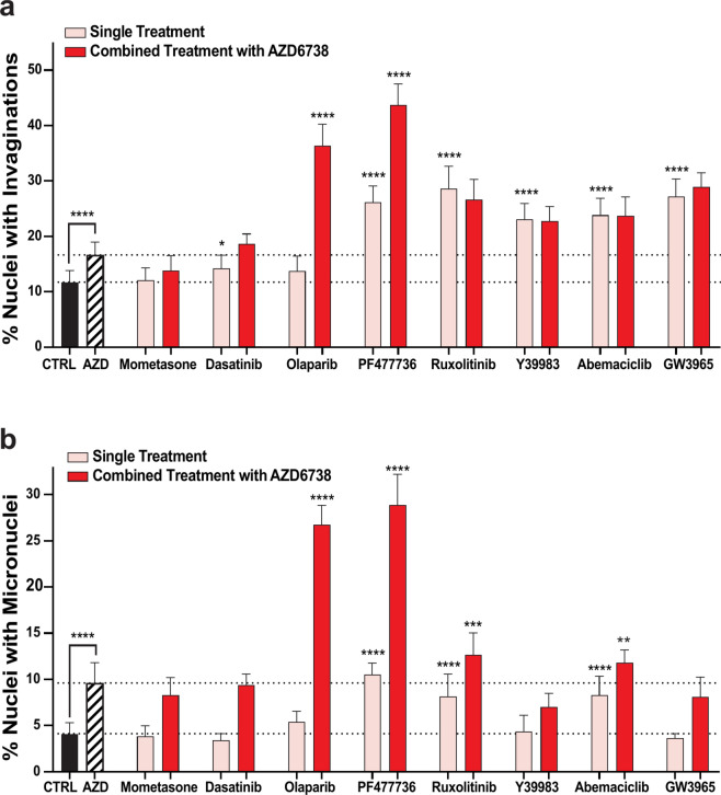
Targeting nuclear mechanics is emerging as a promising therapeutic strategy for sensitizing cancer cells to immunotherapy. Inhibition of the mechano-sensory kinase ATR leads to mechanical vulnerability of cancer cells, causing nuclear envelope softness and collapse and activation of the cGAS-STING-mediated innate immune response. Finding novel compounds that interfere with the non-canonical role of ATR in controlling nuclear mechanics presents an intriguing therapeutic opportunity. We carried out a multiparametric high-content screen to identify small molecules that affect nuclear envelope shape and to uncover novel players that could either ameliorate or further compromise the nuclear mechanical abnormalities of ATR-defective cells. The screen was performed in HeLa cells genetically depleted for ATR. Candidate hits were also tested in combination with the chemical inhibition of ATR by AZD6738, and their efficacy was further validated in the triple-negative breast cancer cell lines BT549 and HCC1937. We show that those compounds enhancing the abnormal nuclear shape of ATR-defective cells also synergize with AZD6738 to boost the expression of interferon-stimulated genes, highlighting the power of multiparametric screens to identify novel combined therapeutic interventions targeting nuclear mechanics for cancer immunotherapy.


Conflict of interest statement

Declarations. Competing interests: The authors declare no competing interests.

Figures

Fig. 1
Fig. 1
Overview of the nuclear morphology image analysis used in the High Content Screening. Representative images of shCTRL and shATR HeLa cells growing in control conditions (DMSO treated) are shown on the left side. Blue: DAPI, Green: Lamin A/C. Nuclei count, roundness and the percent of nuclei with invaginations were estimated by analyzing the DAPI staining. The Lamina Fragmentation was estimated by segmenting the nuclear membrane on the Lamin A/C (FITC staining). The segmentation result on the DAPI channel is shown in the middle panels. NE invaginations are marked in red and the X signs point out nuclei with invaginations. The Lamina segmentation obtained from the FITC channel is shown in black on the right panels. Descriptor vectors of representative images expressed as (nuclei number, roundness, Lamina Fragmentation, percentage of nuclei with invaginations) are for shCTRL (36, 0.97, 1.06, 2.7%) and for shATR (24, 0.94, 1.32, 50%). Scale bar 20 μm.
Fig. 2
Fig. 2
Results of the primary high content screening. Distribution of the z-scores for (a) the percentage of nuclei with invaginations, (b) Lamina Fragmentation and (c) roundness parameters. 18 Assay Plates are represented in the figure. Each compound was tested at three doses (10–1–0.1 μM). Each single dose is distributed in plate A, B and C respectively. Z-scores are calculated for each sample (light gray dots) after normalization on shATR HeLa treated with DMSO (blue dots). shCTRL HeLa cells treated with DMSO are depicted in red. Selected hits (Active-UP/DOWN) are highlighted with green stars according to cut-off value.
Fig. 3
Fig. 3
Analysis of the cellular mechanical properties of HeLa shATR cells treated with ‘worsening’ compounds. (a) Elastic modulus measurements using AFM. Cellular stiffness was measured on shATR HeLa cells treated for 24 h with DMSO or the indicated compounds: Olaparib and Mometasone were tested at 10 μM, Dasatinib at 1 μM. At least 50 cells were detected in three independent experiments. (b) Quantification of nuclear to cytoplasmic YAP signal ratio. shATR HeLa cells were treated with the indicated compounds at two doses for 24 h, then fixed and stained for YAP. The ratio of nuclear and cytoplasmic intensity signals was calculated after background subtraction. Data is reported as mean ± SD of three independent experiments with at least 3 technical replicates for the treated samples and at least 12 technical replicates for the controls in each experiment. **p ≤ 0.01, ****p ≤ 0.0001 by ordinary one-way ANOVA test using Bonferroni’s correction for multiple comparisons. (c) Immunofluorescence images showing YAP cellular distribution under the indicated conditions. Scale bar 20 μm.
Fig. 4
Fig. 4
Effects of the ‘worsening’ compounds combined with AZD6738 on NE invaginations and MN in HeLa cells. HeLa cells were treated with AZD6738 2 μM. 24 h later, the selected compounds were added as single agents (pink bars) or in combination with AZD6738 (red bars) for further 24 h. All the compounds were used at 10uM with the exception of Dasatinib and PF-477736 that were tested at the dose of 1 μM. Doses were selected considering those that impact nuclear morphology without inducing a reduction in the number of nuclei by more than 35%. The effect of the compounds on NE invaginations (a) or micronuclei (b) is shown in the bar graphs. Data are reported as mean ± SD of three biological replicates with at least 3 technical replicates for the treated samples and at least 9 technical replicates for the DMSO conditions. Statistical analysis by ordinary one-way ANOVA test using Bonferroni’s correction for multiple comparisons was applied, *p ≤ 0.05, **p ≤ 0.01, ***p ≤ 0.001, ****p ≤ 0.0001.
Fig. 5
Fig. 5
Effect of the 8 ‘worsening’ compounds on the nuclear morphometric parameters of human TNBC cell lines. Each molecule was tested at two doses for 24 h on HCC1937 cells (a) or BT549 cells (b). Doses were selected considering those that did not reduce the count of nuclei by more than 35% for the respective cell line. Data are reported as mean ± SD of two independent experiments with at least 3 technical replicates for the treated samples and at least 6 technical replicates for the DMSO conditions in each experiment. Statistical analysis by ordinary one-way ANOVA test using Bonferroni’s correction for multiple comparisons was applied, *p ≤ 0.05, **p ≤ 0.01, ***p ≤ 0.001, ****p ≤ 0.0001.
Fig. 6
Fig. 6
Effects of combined treatments with AZD6738 on NE invaginations and MN in the TNBC cell line HCC1937. Combined treatments are reported in red, while single treatments are in pink. Data are reported as mean ± SD of three independent experiments with at least 3 technical replicates for the treated samples and at least 9 technical replicates for the DMSO conditions in each experiment. All the compounds were tested at 10 μM, except PF-477736 and Dasatinib tested at 1 μM and 0.1 μM respectively. AZD6738 was tested at 3 μM. Doses were selected considering those that impact nuclear morphology without inducing a reduction in the nuclei count greater than 35%. The effect of the compounds on NE invaginations (a) or micronuclei (b) is shown in the bar graphs. Statistical analysis by ordinary one-way ANOVA test using Bonferroni’s correction for multiple comparisons was applied, **p ≤ 0.01, ***p ≤ 0.001, ****p ≤ 0.0001.
Fig. 7
Fig. 7
The ‘worsening’ compounds stimulate a cGAS-STING-mediated inflammatory ISG response in HCC1937 cells when combined with AZD6738. Heat-map graph showing the induction of a selected panel of ISG genes normalized to the DMSO condition in HCC1937 cells treated with ATRi AZD6738 3 μM for 48 h; AZD6738 was administered alone or in combination with the ‘worsening’ compounds (24 h treatment). Mometasone and Olaparib were tested at 10 μM, while PF-477736 and Dasatinib were tested at 1 μM and 0.1 μM respectively. Bar graphs show the ISG induction levels as an average of three independent biological replicates (left panel). Statistical analysis by ordinary one-way ANOVA test using Bonferroni’s correction for multiple was applied, *p ≤ 0.05, **p ≤ 0.01, ***p ≤ 0.001.

Similar articles

References

    1. Mekhail, K. & Moazed, D. The nuclear envelope in genome organization, expression and stability. Nat. Rev. Mol. Cell Biol.11, 317–328. 10.1038/nrm2894 (2010). - PMC - PubMed
    1. Bastianello, G. & Foiani, M. Mechanisms controlling the mechanical properties of the nuclei. Curr. Opin. Cell Biol.84, 102222. 10.1016/j.ceb.2023.102222 (2023). - PubMed
    1. Nader, G. P. F., Williart, A. & Piel, M. Nuclear deformations, from signaling to perturbation and damage. Curr. Opin. Cell Biol.72, 137–145. 10.1016/j.ceb.2021.07.008 (2021). - PubMed
    1. Isermann, P. & Lammerding, J. Nuclear mechanics and mechanotransduction in health and disease. Curr. Biol.23, R1113-1121. 10.1016/j.cub.2013.11.009 (2013). - PMC - PubMed
    1. Kidiyoor, G. R. et al. ATR is essential for preservation of cell mechanics and nuclear integrity during interstitial migration. Nat. Commun.11, 4828. 10.1038/s41467-020-18580-9 (2020). - PMC - PubMed

LinkOut - more resources