. 2024 Dec 28;14(1):30876.
doi: 10.1038/s41598-024-81668-5.

Quinoline-thiosemicarbazone-1,2,3-triazole-acetamide derivatives as new potent α-glucosidase inhibitors

Affiliations

Quinoline-thiosemicarbazone-1,2,3-triazole-acetamide derivatives as new potent α-glucosidase inhibitors

Aynaz Khademian et al. Sci Rep. .

Abstract

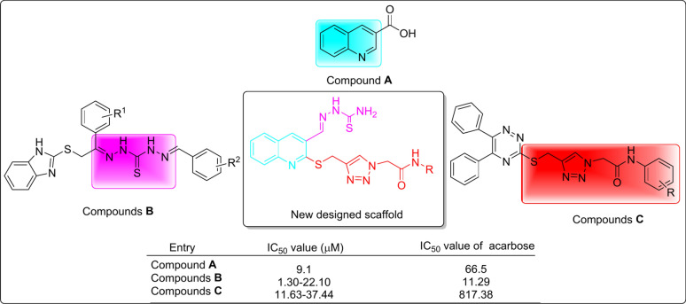
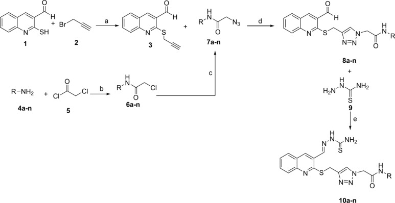
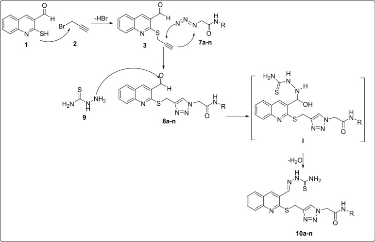
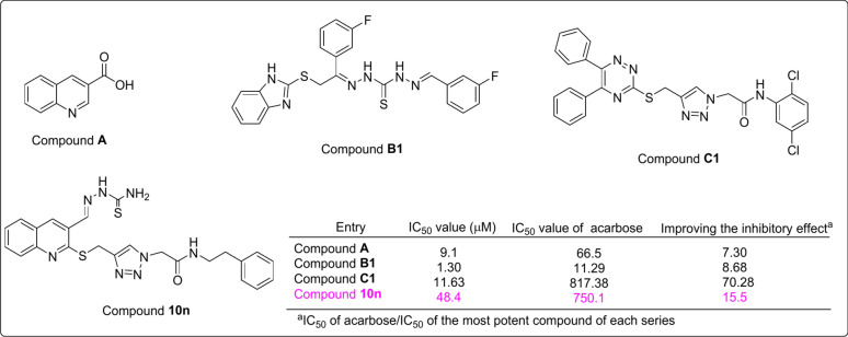
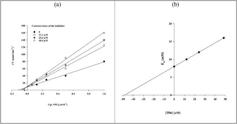
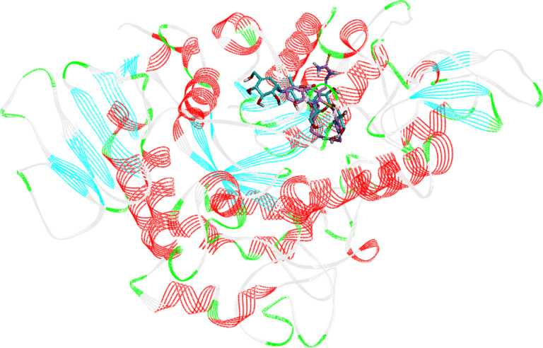
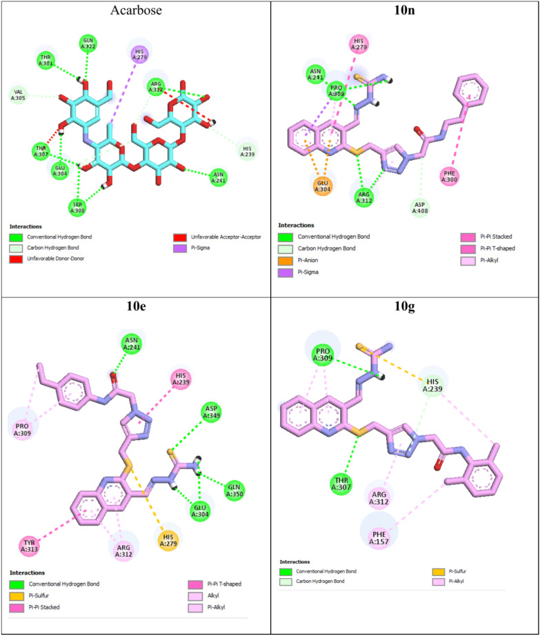
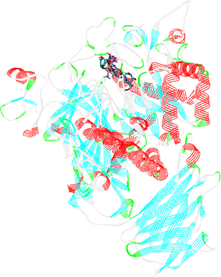
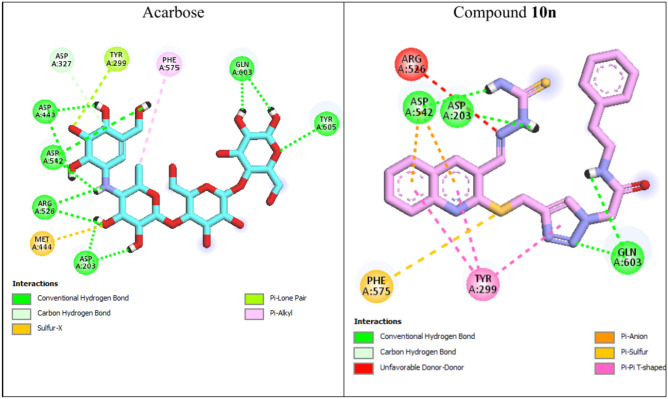
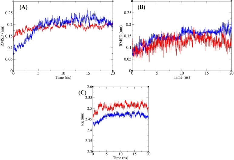
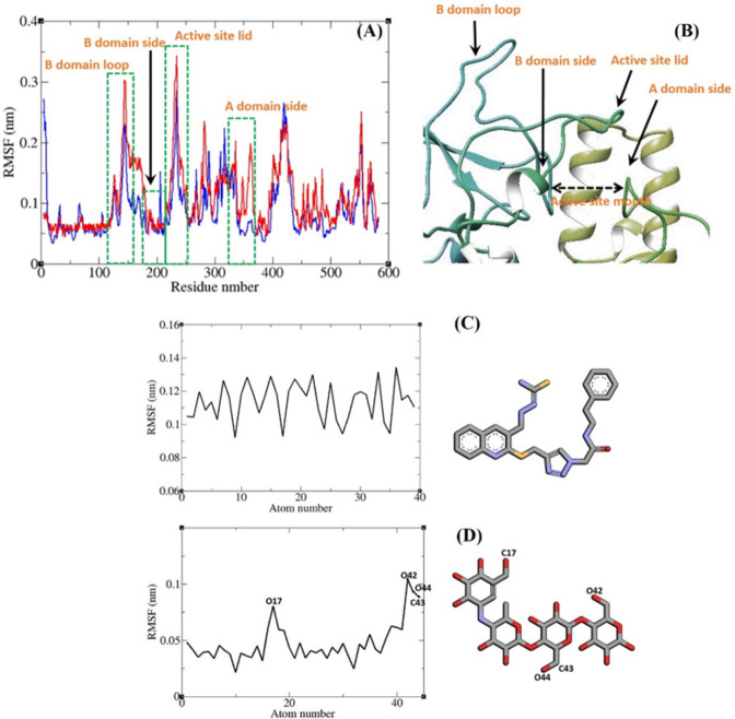
In this work, a novel series of quinoline-thiosemicarbazone-1,2,3-triazole-aceamide derivatives 10a-n as new potent α-glucosidase inhibitors was designed, synthesized, and evaluated. All the synthesized derivatives 10a-n were more potent than acarbose (positive control). Representatively, (E)-2-(4-(((3-((2-Carbamothioylhydrazineylidene)methyl)quinolin-2-yl)thio)methyl)-1H-1,2,3-triazol-1-yl)-N-phenethylacetamide (10n), as the most potent entry, with IC50 = 48.4 µM was 15.5-times more potent than acarbose. According to kinetic study, compound 10n was a competitive inhibitor against α-glucosidase. This compound formed the desired interactions with important residues of the binding pocket of α-glucosidase with favorable binding energy in the molecular docking and molecular dynamics. Compounds 10n, 10e, and 10 g as the most potent compounds among the synthesized compounds were evaluated in term of pharmacokinetics and toxicity via online servers. These evaluations predicted that compounds 10n, 10e, and 10 g had good pharmacokinetic properties and toxicity profile.

Keywords: 1,2,3-Triazole; Quinoline; Thiosemicarbazone; α-Glucosidase inhibitors.


Conflict of interest statement

Declarations. Competing interests: The authors declare no competing interests. Ethics approval and consent to participate: Not applicable. Consent for publication: Not applicable.

Figures

Fig. 1
Fig. 1
Structures and inhibitory activities of the reported compounds A-C that were used in the design of quinoline-thiosemicarbazone-1,2,3-triazole-aceamide skeleton as a new scaffold with anti-α-glucosidase property.
Fig. 2
Fig. 2
Synthesis of quinoline-thiosemicarbazone-1,2,3-triazole-aceamide derivatives 10a-n: (a) K2CO3, Aceton, RT, 1 h; (b) DMF, RT, 1 h; (c) Et3N, H2O/t-BuOH, RT, 1 h; (d) CuSO4·5H2O, Sodium ascorbate, RT, 24–48 h; (e) Methanol, Acetic acid, Reflux, 4 h.
Fig. 3
Fig. 3
The proposed reaction mechanism for the formation of quinoline-thiosemicarbazone-1,2,3-triazole-aceamide derivatives 10a-n.
Fig. 4
Fig. 4
Diagram of SAR of compounds 10a-m against α-glucosidase.
Fig. 5
Fig. 5
Comparison of α-glucosidase inhibitory activity between template compounds A, B1 and C1 with the most potent new compound 10n.
Fig. 6
Fig. 6
(a) Lineweaver–Burk plot for compound 10n (the concentrations of compound 10n were 0, 12.1, 24.2, and 48.4 µM. (b) Secondary re-plot of slopes of Lineweaver–Burk plot vs various concentrations of the compound 10n.
Fig. 7
Fig. 7
Acarbose (cyan) and the most potent compound 10n (pink) superimposed in the α-glucosidase active site.
Fig. 8
Fig. 8
Interaction modes of acarbose and the most potent compounds 10n, 10e, and 10 g in the active site of α-glucosidase.
Fig. 9
Fig. 9
Superimposed structure of acarbose (cyan) and the most potent compound 10n (pink) in the human α-glucosidase active site.
Fig. 10
Fig. 10
Interaction modes of acarbose and the most potent compound 10n in the active site of human α-glucosidase.
Fig. 11
Fig. 11
Superimposed RMSD of Cα atoms of α-glucosidase in complex with 10n (blue) and acarbose (red) (A). Superimposed RMSD of 10n (blue) and acarbose (red) in complex with α-glucosidase (B). Time dependence of the radius of gyration (Rg) graph of α-glucosidase in complex with 10n (blue) and acarbose (red) (C).
Fig. 12
Fig. 12
(A) RMSF graph of the Cα atoms of α-glucosidase in complex with 10n (blue) and acarbose (red). Close-up representation of α-glucosidase active site (B). RMSF graph of the heavy atoms of 10n (C) and acarbose (D) in complex with α-glucosidase. Structure of these compounds are illustrated.

References

    1. Roglic, G. WHO Global report on diabetes: A summary. Int. J. Noncommun. Dis.1, 3–8 (2016).
    1. Dabelea, D. et al. Prevalence of type 1 and type 2 diabetes among children and adolescents from 2001 to 2009. Jama311, 1778–1786 (2014). - PMC - PubMed
    1. Kulkarni, A., Thool, A. R. & Daigavane, S. Understanding the clinical relationship between diabetic retinopathy, nephropathy, and neuropathy: A comprehensive review. Cureus16, e56674 (2024). - PMC - PubMed
    1. Kim, Y. M., Jeong, Y. K., Wang, M. H., Lee, W. Y. & Rhee, H. I. Inhibitory effect of pine extract on α-glucosidase activity and postprandial hyperglycemia. Nutrition21, 756–761 (2005). - PubMed
    1. Choi, C. W. et al. Yeast α-glucosidase inhibition by isoflavones from plants of Leguminosae as an in vitro alternative to acarbose. J. Agric. Food Chem.58, 9988–9993 (2010). - PubMed

MeSH terms

LinkOut - more resources