. 2024 Dec 28;14(1):30879.
doi: 10.1038/s41598-024-81675-6.

Generation and characterization of CRISPR-Cas9-mediated XPC gene knockout in human skin cells

Affiliations

Generation and characterization of CRISPR-Cas9-mediated XPC gene knockout in human skin cells

Ali Nasrallah et al. Sci Rep. .

Abstract

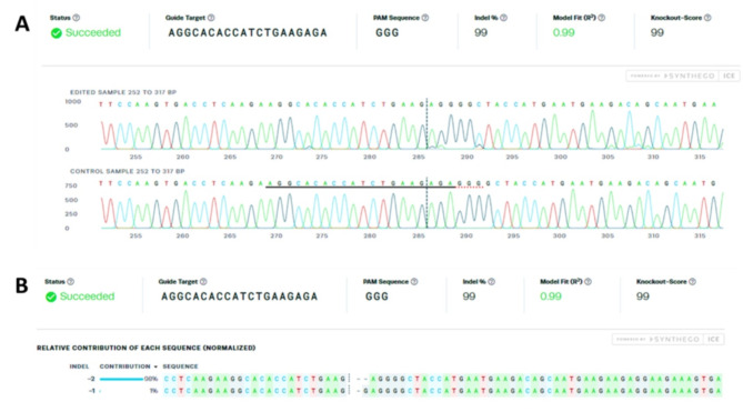
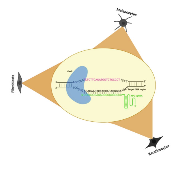
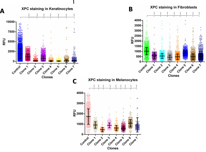
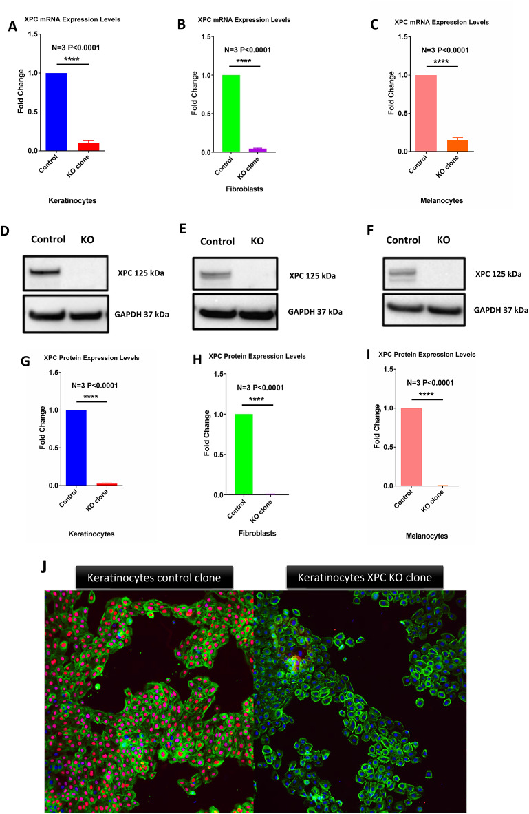
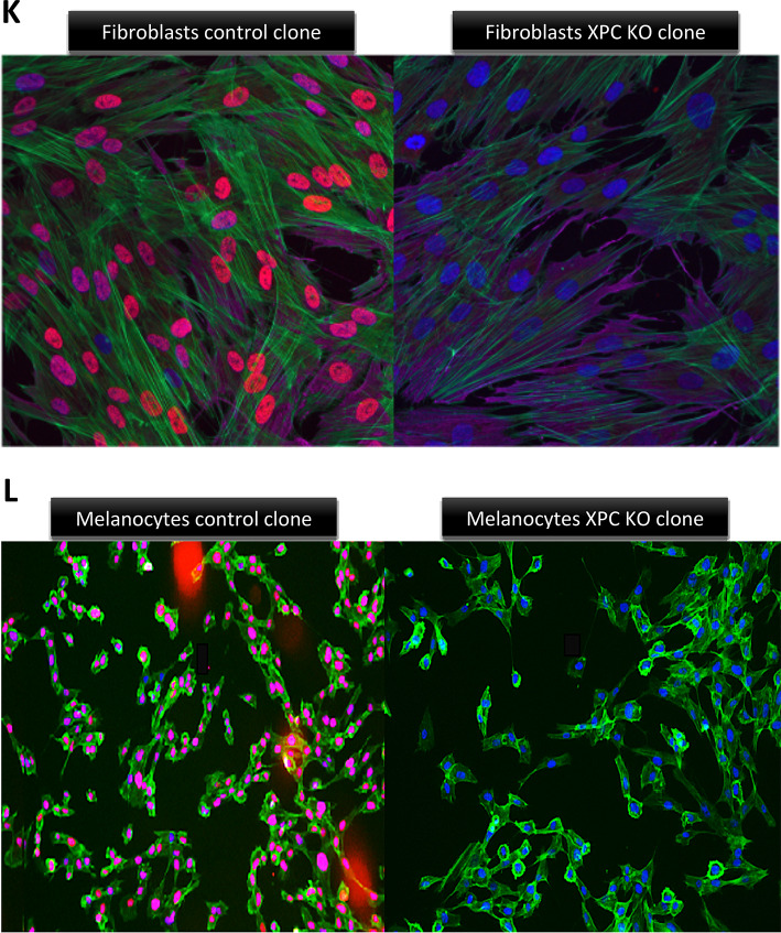
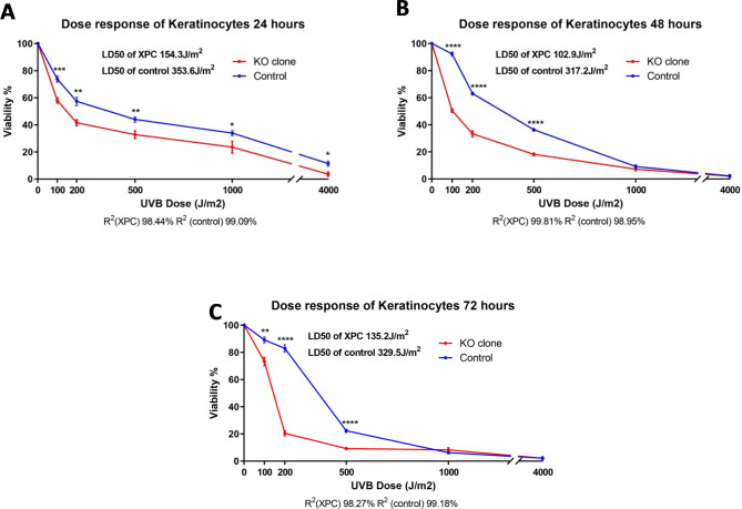
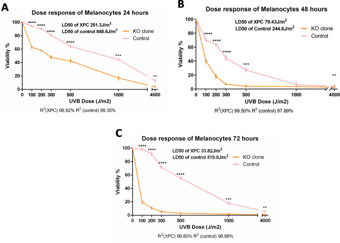
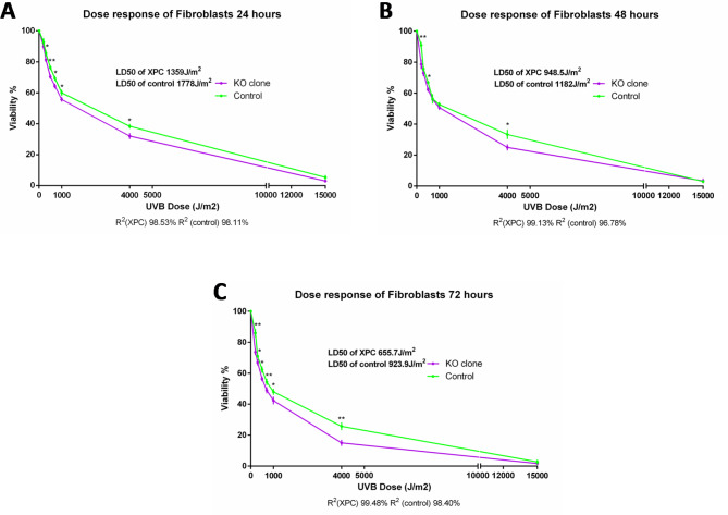
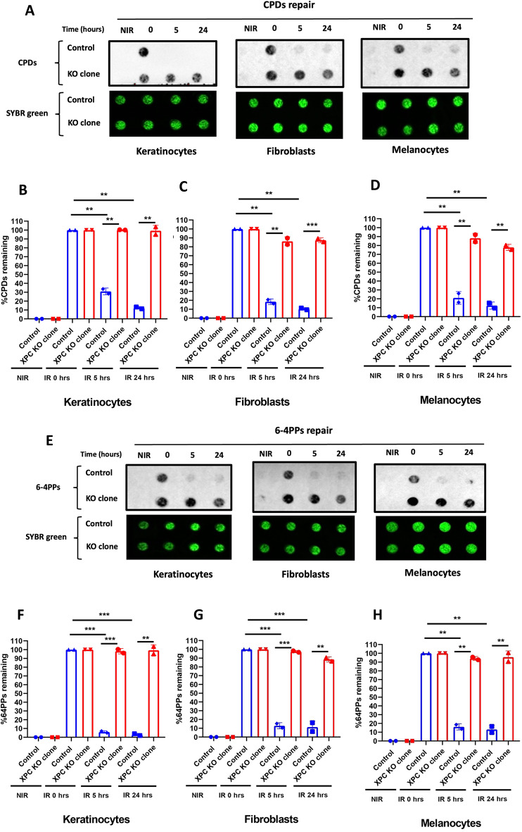
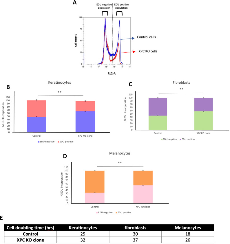
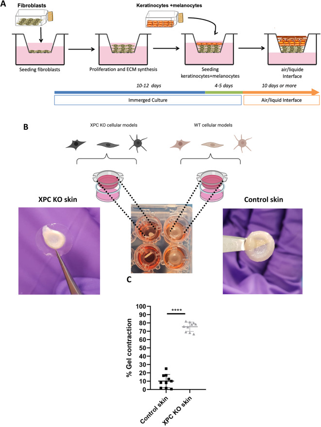
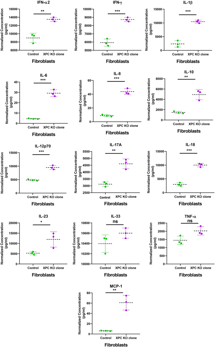
Xeroderma pigmentosum group C (XPC) is a versatile protein crucial for sensing DNA damage in the global genome nucleotide excision repair (GG-NER) pathway. This pathway is vital for mammalian cells, acting as their essential approach for repairing DNA lesions stemming from interactions with environmental factors, such as exposure to ultraviolet (UV) radiation from the sun. Loss-of-function mutations in the XPC gene confer a photosensitive phenotype in XP-C patients, resulting in the accumulation of unrepaired UV-induced DNA damage. This remarkable increase in DNA damage tends to elevate by 10,000-fold the risk of developing melanoma and non-melanoma skin cancers. To date, creating accurate and reproducible models to study human XP-C disease has been an important challenge. To tackle this, we used CRISPR-Cas9 technology in order to knockout the XPC gene in various human skin cells (keratinocytes, fibroblasts, and melanocytes). After validation of the knockout in these edited skin cells, we showed that they recapitulate the major phenotypes of XPC mutations: photosensitivity and the impairment of UV-induced DNA damage repair. Moreover, these knockout cells demonstrated a reduced proliferative capacity compared to their respective controls. Finally, to better mimic the disease environment, we built a 3D reconstructed skin using these XPC knockout skin cells. This model exhibited an abnormal behavior, showing an extensive remodeling of its extracellular matrix compared to normal skin. Analyzing the composition of the fibroblast secretome revealed a significant augmented shift in the inflammatory response following XPC knockout. Our innovative "disease on a dish" approach can provide valuable insights into the molecular mechanisms underlying XP-C disease, paving the way to design novel preventive and therapeutic strategies to alleviate the disease phenotype. Also, given the high risk of skin cancer onset in XP-C disease, our new approach can serve as a link to draw novel insights into this elusive field.

Keywords: CRISPR-Cas9; DNA damage; Skin; UV irradiation; XP-C disease.


Conflict of interest statement

Declarations. Competing interests: The authors declare no competing interests.

Figures

Fig. 1
Fig. 1
Sequencing analysis of the N/TERT-2G XPC knockout (KO) heterogeneous population compared to control cells. (A,B) The N/TERT-2G XPC knockout (KO) heterogeneous population subjected to Sanger sequencing and compared to the control DNA sequence. (A) The sgRNA target region, the PAM sequence, and the knockout score are highlighted. Comparison of both sequences showed a predominant two-nucleotide (AG) indel mutation, in the exon 3 site. (B) The percentage of distribution of edits (indels) in the DNA sequence of the N/TERT-2G XPC knockout (KO) heterogeneous population.
Fig. 2
Fig. 2
Schematic representation of the XPC gene target site for editing in human immortalized keratinocytes, fibroblasts, and melanocytes.
Fig. 3
Fig. 3
(AC) Selection of the XPC gene homozygous knockout (KO) clones in N/TERT-2G, S1F/TERT-1 and Mel-ST cell lines. (A) Selection of the XPC gene homozygous knockout (KO) clones in the N/TERT-2G cell line. Five edited N/TERT-2G clones (2,4,5,6, and 7) showed an absence of XPC’s relative fluorescence unit (RFU) compared to their control cells. (B) Selection of the XPC gene homozygous knockout (KO) clones in S1F/TERT-1 cell line. Six edited S1F/TERT-1 clones (1,2,3,4,6, and 7) showed an absence of XPC’s relative fluorescence unit (RFU) compared to their control cells. (C) Selection of the XPC gene homozygous knockout (KO) clones in Mel-ST cell line. Six edited Mel-ST clones (1,2,3,4,5, and 7) showed an absence of XPC’s relative fluorescence unit (RFU) compared to their control cells. ***p-value < 0.001 unpaired Student T test.
Fig. 4
Fig. 4
Validation of XPC gene knockout (KO) in N/TERT-2G, S1F/TERT-1 and Mel-ST cell lines. (AC) RT-qPCR analysis shows the absence of XPC mRNA in knockout clones compared to their associated controls (****p < 0.0001). (DI) Western blot confirms the lack of XPC protein in knockout clones across all cell types, with GAPDH as a loading control (****p < 0.0001). (JL) Immunofluorescence staining reveals no XPC protein in the nuclei of knockout cells, while the control cells show XPC presence (in red). Cropped blots are shown; full-length blots are in Supplementary Figure (S4D–F).
Fig. 4
Fig. 4
Validation of XPC gene knockout (KO) in N/TERT-2G, S1F/TERT-1 and Mel-ST cell lines. (AC) RT-qPCR analysis shows the absence of XPC mRNA in knockout clones compared to their associated controls (****p < 0.0001). (DI) Western blot confirms the lack of XPC protein in knockout clones across all cell types, with GAPDH as a loading control (****p < 0.0001). (JL) Immunofluorescence staining reveals no XPC protein in the nuclei of knockout cells, while the control cells show XPC presence (in red). Cropped blots are shown; full-length blots are in Supplementary Figure (S4D–F).
Fig. 5
Fig. 5
XPC-KO N/TERT-2G keratinocyte cells manifest increased hypersensitivity to UVB irradiation in a dose- and time-dependent manner. (AC) Both XPC knockout (KO) and control cells were exposed to various doses of UVB, and their viability was assessed after 24 (A), 48 (B), and 72 (C) hours using the trypan blue exclusion test. XPC KO cells demonstrated a notably steeper and statistically significant reduction in viability with rising UVB doses compared to control cells. Viability was determined as a percentage of the control, with non-irradiated cells representing 100% viability. Statistical analysis revealed a highly significant difference: *p-value < 0.05, **p-value < 0.01, ***p-value < 0.001, ****p-value < 0.0001 (unpaired t-test). The reported results are the average of three separate biological experiments (N = 3).
Fig. 6
Fig. 6
XPC KO Mel-ST melanocyte cells manifest increased hypersensitivity to UVB irradiation in a dose and time dependent manner. (A) Both XPC knockout (KO) and control cells were exposed to various doses of UVB, and their viability was assessed after 24 (A), 48 (B), and 72 (C) hours using the trypan blue exclusion test. XPC KO cells exhibited a more pronounced and statistically significant reduction in viability as the UVB dose increased, in contrast to control cells. Viability was determined by calculating the percentage in relation to the control, where non-irradiated cells represented 100% viability. The statistical significance was denoted as **p-value < 0.01, ***p-value < 0.001, ****p-value < 0.0001 (unpaired t-test). The findings are based on the average of three independent biological replicates (N = 3).
Fig. 7
Fig. 7
XPC KO S1F/TERT-1 fibroblast cells manifest a slight hypersensitivity to UVB irradiation in a dose- and time-dependent manner. (A) Both XPC knockout (KO) and control cells were exposed to various doses of UVB, and their viability was assessed after 24 (A), 48 (B), and 72 (C) hours using trypan blue exclusion test. XPC KO cells exhibited a slight statistically significant reduction in viability as the UVB dose increased, as compared to the control cells. Viability was determined by calculating the percentage in relation to the control, where non-irradiated cells were considered to have 100% viability. The statistical significance was indicated as *p-value < 0.05, **p-value < 0.01 (unpaired t-test). The presented results are the average of three independent biological replicates (N = 3).
Fig. 8
Fig. 8
XPC KO N/TERT-2G keratinocyte, S1F/TERT-1 fibroblast, and Mel-ST melanocyte cells manifest a significantly persistent and unrepaired 6-4PPs and CPDs after UVB irradiation. (AH) Repair kinetics of CPDs and 6-4PPs were evaluated by immunodot blot and compared between control and XPC KO keratinocytes, fibroblasts, and melanocytes. Immuno-dot blots show that the removal kinetics of CPDs (top panel A) and 6-4PPs (bottom panel E) are significantly decreased in XPC KO keratinocytes (A), fibroblasts (B), and melanocytes (C) compared to their respective control cells. SYBR green was used as a loading control. (BD,FH) percentage of remaining CPDs and 6-4PPs at indicated time points after irradiation was calculated by comparison with the initial levels in keratinocytes (B,F), fibroblasts (C,G), and melanocytes (D,H). **p-value < 0.01, ***p-value < 0.001 unpaired t-test. NIR: non-irradiated The reported results are the average of two separate biological experiments (N = 2).
Fig. 9
Fig. 9
XPC KO manifests a partial halting in the proliferative capacity of N/TERT-2G keratinocytes, S1F/TERT-1 fibroblasts, and Mel-ST melanocytes. (AD) EdU incorporation assay for XPC KO and control keratinocytes, fibroblasts, and melanocytes. EdU was added to the cell media for 5 h. EDU incorporation was measured using flow cytometry. A representative flow-cytometry plot (A) and quantification (BD) of EdU incorporation are shown **p-value < 0.01 (unpaired t-test). (E) Representation of the population doubling time in hours for XPC KO clones and their associated control cells.
Fig. 10
Fig. 10
Shrinkage of the fibrin gel in the XPC KO skin model during differentiation processes. (A) The protocol consists of seeding separately control XPC KO fibroblasts in a specific gel termed fibrin, in insert support to permit the formation of the dermal equivalent. Afterwards, XPC KO and control melanocytes and keratinocytes are seeded on top of either the XPC KO or control dermal equivalent and allowed to proliferate and differentiate. (B) An extensive shrinkage of scaffold was observed in reconstructions with XPC KO cells. Gel diameters from 10 XPC KO versus 10 control reconstructed skins were measured using ImageJ software. (C) The percentage of gel contraction was calculated using the formula 100x (well diameter-gel diameter)/well diameter. **** p < 0.0001 unpaired t-test.
Fig. 11
Fig. 11
XPC KO induces a rise in the inflammatory secretome signature secreted by S1F/TERT-1 fibroblasts. Knocking out XPC in S1F/TERT-1 fibroblasts induces an increase in the human inflammatory secretion profile, which encompasses both chemokines and cytokines, including a rise in IL-1β, IFN-α2, IFN-γ, TNF, MCP-1 (CCL2), IL-6, IL-8 (CXCL8), IL-10, IL-12p70, IL-17 A, IL-18, IL-23, and IL-33. Subsequently, samples were acquired using a FACSCanto™ II cytometer and analyzed utilizing the online QOGNIT LEGENDplex™ program. The statistical significance was denoted as **p-value < 0.01, ***p-value < 0.001, ****p-value < 0.0001 (unpaired t-test). The results presented are the mean of three biological replicates (N = 3).

Similar articles

Cited by

References

    1. Griffiths, H. R., Mistry, P., Herbert, K. E. & Lunec, J. Molecular and cellular effects of ultraviolet light-induced genotoxicity. Crit. Rev. Clin. Lab. Sci.35, 189–237 (1998). - PubMed
    1. Douki, T. The variety of UV-induced pyrimidine dimeric photoproducts in DNA as shown by chromatographic quantification methods. Photochem. Photobiol Sci. Off J. Eur. Photochem. Assoc. Eur. Soc. Photobiol. 12, 1286–1302 (2013). - PubMed
    1. Hung, K. F., Sidorova, J. M., Nghiem, P. & Kawasumi, M. The 6 – 4 photoproduct is the trigger of UV-induced replication blockage and ATR activation. Proc. Natl. Acad. Sci.117, 12806–12816 (2020). - PMC - PubMed
    1. Sakamoto, A. et al. Immunoexpression of ultraviolet photoproducts and p53 mutation analysis in atypical fibroxanthoma and superficial malignant fibrous histiocytoma. Mod. Pathol. Off J. U S Can. Acad. Pathol. Inc. 14, 581–588 (2001). - PubMed
    1. Nasrallah, A. et al. Xeroderma Pigmentosum C: A Valuable Tool to Decipher the Signaling Pathways in Skin Cancers. Oxid. Med. Cell. Longev. 6689403 (2021). - PMC - PubMed

LinkOut - more resources