. 2024 Dec 28;14(1):31008.
doi: 10.1038/s41598-024-81998-4.

Viral coat proteins decrease the gene silencing activity of cognate and heterologous viral suppressors

Affiliations

Viral coat proteins decrease the gene silencing activity of cognate and heterologous viral suppressors

Dóra Pinczés et al. Sci Rep. .

Abstract

Plant viruses have evolved different viral suppressors of RNA silencing (VSRs) to counteract RNA silencing which is a small RNA-mediated sequence-specific RNA degradation mechanism. Previous studies have already shown that the coat protein (CP) of cucumber mosaic virus (CMV) reduced RNA silencing suppression (RSS) activity of the VSR of CMV, the 2b protein. To demonstrate the universality of this CP-VSR interference, our study included three different viruses: CMV and peanut stunt virus (PSV) from the Bromoviridae, and plum pox virus (PPV) from the Potyviridae family. The RSS activity of the three VSRs (CMV 2b, PSV 2b, and PPV HC-Pro) was compared using Agrobacterium-mediated transient expression in Nicotiana benthamiana and the effect of CMV CP, PSV CP and PPV CP was validated on the RSS activity of their cognate and heterologous VSRs as well. Furthermore, the VSRs were also evaluated in PTGS suppressor-deficient CMV mutant (CMV NVE/10-12/AAA) virus-infected plants. The joint presence of CPs and VSRs resulted in decreased RSS activity in each combination, regardless of the origin of the two proteins, suggesting a universal role of the viral CPs in fine tuning of RSS. Interestingly the PSV CP elicited the strongest negative effect on the RSS activity of all three VSRs.


Conflict of interest statement

Declarations. Competing interests: The authors declare no competing interests.

Figures
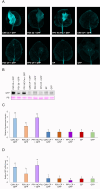
Fig. 1
Fig. 1
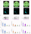
Comparison of the RSS activity of CMV, PSV and PPV proteins in patch assays. (A) Agroinfiltration assay of the VSRs and CPs of CMV, PSV and PPV on N. benthamiana leaves. Agrobacterium expressing GFP was coinfiltrated with either the VSRs or the CPs of CMV, PSV and PPV and visualized 5 days post agroinfiltration (dpa). Agrobacterium carrying an empty plasmid (EP) and GFP were used as negative controls. (B) Western blot with anti-GFP antibody was used to detect the expressed GFP on the agroinfiltrated patches. Ponceau staining (PS) served as loading control. Original blots are presented in Supplementary Fig. S11. (C) The relative fluorescence of the infiltrated patches was measured using iBright Analysis Software. The fluorescence of the degraded GFP-expressing patch collected 5 dpa was set as 1.0. Significant differences were observed using Tukey’s multiple comparison test (*P < 0.05). The different letters above the bars indicate significant difference in fluorescence intensity among the different constructs. (D) RT-qPCR was used to detect the mRNA levels of GFP. The mRNA of N. benthamiana EF1α was used as internal control. Significant differences were observed using Tukey’s multiple comparison test (*P < 0.05). The different letters above the bars indicate significant difference in GFP mRNA level among the different constructs.
Fig. 2
Fig. 2
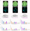
Analysis of the RSS activity of CMV 2b, PSV 2b, and PPV HC-Pro coexpressed with cognate CPs. (A) Coinfiltration assay of CMV 2b plus CMV CP, PSV 2b plus PSV CP and PPV HC-Pro plus PPV CP and GFP on N. benthamiana leaves. Agrobacterium carrying the CPs and GFP were used as controls. (B) Western blot with anti-GFP antibody was used to detect the expressed GFP in the agroinfiltrated patches. For the detection of the VSRs, anti-CMV 2b, anti-HIS and anti-FLAG antibodies were used. For the detection of the CPs, anti-Cucumo CP and anti-PPV CP antibodies were used. Ponceau staining (PS) served as loading control. Original blots are presented in Supplementary Fig. S12-14. (C) The relative fluorescence of the infiltrated patches was measured using iBright Analysis Software. The fluorescence of the degraded GFP-expressing patch collected 5 dpa was set as 1.0. Significant differences were observed using one-way ANOVA model and Games-Howell post hoc test (Supplementary Fig. S3) (*P < 0.05). The different letters above the bars indicate significant difference among isolates. (D) RT-qPCR was used to detect the mRNA levels of GFP. The mRNA of N. benthamiana EF1α was used as internal control. Significant differences were observed using one-way ANOVA model and Games-Howell post hoc test (Supplementary Fig. S4) (*P < 0.05). The different letters above the bars indicate a significant difference among isolates.
Fig. 3
Fig. 3
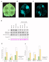
Effect of the different CPs on the RSS activity of CMV 2b, PSV 2b and PPV HC-Pro suppressor proteins, in mixed combinations. (A) Coinfiltration assay of VSRs and CPs of CMV, PSV and PPV on N. benthamiana leaves. Agrobacterium carrying the CPs and GFP were used as controls. (B) Western blot analysis with anti-GFP antibody was used to detect the expressed GFP on the agroinfiltrated patches. For the detection of the CMV, PSV, PPV VSRs and CPs, anti-CMV 2b, anti-HIS, anti-FLAG, anti-Cucumo CP and anti-PPV CP antibodies were used. Ponceau staining (PS) served as loading control. Original blots are presented in Supplementary Fig. S15-17. (C) The relative fluorescence of the infiltrated patches was measured using iBright Analysis Software. The fluorescence of the degraded GFP-expressing patch collected 5 dpa was set as 1.0. Significant differences were observed using Tukey’s multiple comparison test and Games-Howell Post Hoc test (*P < 0.05) (Supplementary Fig. S5). The different letters above the bars indicate significant difference among isolates. (D) RT-qPCR was used to detect the mRNA levels of GFP. The mRNA of N. benthamiana EF1α was used as internal control. Significant differences were observed using Tukey’s multiple comparison test and Games-Howell Post Hoc test (*P < 0.05) (Supplementary Fig. S6). The different letters above the bars indicate significant difference among isolates.
Fig. 4
Fig. 4
Effect of CMV infection on the RSS activity of CMV 2b, PSV 2b and PPV HC-Pro suppressor proteins (A) Schematic representation of the agroinfiltrated patches on N. benthamiana leaves (B) Infiltration assay of VSRs of CMV, PSV and PPV on non-infected N. benthamiana leaves. Agrobacterium expressing GFP was used as control. (C) Infiltration assay of VSRs of CMV, PSV and PPV on NVE/10–12/AAA CMV infected N. benthamiana leaves. Agrobacterium expressing GFP was used as a control. (D) Western blot analysis with anti-GFP antibody was used to detect the expressed GFP on the agroinfiltrated patches. For the detection of the CMV 2b and PSV 2b anti-HIS and for PPV HC-Pro detection anti-FLAG antibodies were used. The arrows and the asterisks indicate the specific and non-specific bands, respectively. Ponceau staining (PS) served as loading control. Blots cropped from different gels were marked with black lines. Original blots are presented in Supplementary Fig. S18-20. (E) The relative fluorescence of the infiltrated patches was measured using iBright Analysis Software. The fluorescence intensity of the infiltrated patches was normalized to GFP fluorescence. Significant differences were observed using Student’s two tailed t-test (*P < 0.05) (Supplementary Fig. S7A). Asterisks indicate the statistically significant difference between non inoculated and CMV NVE/10–12/AAA inoculated N. benthamiana when infiltrated with the same VSR (F) RT-qPCR was used to detect the mRNA levels of GFP. The mRNA of N. benthamiana EF1α was used as internal control. Significant differences were observed using Student’s two tailed t-test (*P < 0.05) (Supplementary Fig. S7B). Asterisks indicate the statistically significant difference between non inoculated and CMV NVE/10–12/AAA inoculated N. benthamiana when infiltrated with the same VSR.
Fig. 5
Fig. 5
Quantification of plant gene expressions involved in gene silencing mechanism. The expression level of various Nicotiana benthamiana genes involved in post transcriptional gene silencing (AGO1, AGO5, RDR6, DCL4), in the presence of CMV 2b, PSV 2b and PPV HC-Pro infiltrated alone or coinfiltrated with CMV CP. GFP and CMV CP single infiltrations were used as controls. Statistically significant differences were observed using Tukey’s multiple comparison test and Games-Howell post hoc test (*P < 0.05). The different letters above the bars indicate significant differences between groups.

Similar articles

References

    1. Waterhouse, P. M., Wang, M.-B. & Lough, T. Gene silencing as an adaptive defence against viruses. Nature411, 834–842 (2001). - PubMed
    1. Meister, G. & Tuschl, T. Mechanisms of gene silencing by double-stranded RNA. Nature431, 343–349 (2004). - PubMed
    1. Vance, V. & Vaucheret, H. RNA silencing in plants-defense and counterdefense. Science292, 2277–2280 (2001). - PubMed
    1. Li, W. X. & Ding, S. W. Viral suppressors of RNA silencing. Curr. Opin. Biotechnol.12, 150–154 (2001). - PubMed
    1. Burgyán, J. & Havelda, Z. Viral suppressors of RNA silencing. Trends Plant Sci.16, 265–272 (2011). - PubMed

Substances

LinkOut - more resources