. 2024 Dec 28;14(1):31019.
doi: 10.1038/s41598-024-82176-2.

Exploring the role of the MUC1 mucin in human oral lubrication by tribological in vitro studies

Affiliations

Exploring the role of the MUC1 mucin in human oral lubrication by tribological in vitro studies

Ianis Ammam et al. Sci Rep. .

Abstract

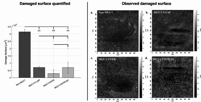
In the context of the oral cavity, an organic layer known as the mucosal pellicle (MP) adheres to the surface of the oral epithelium, playing a pivotal role in lubricating and safeguarding oral tissues. The formation of the MP is driven by interactions between a transmembrane mucin known as MUC1, located on the oral epithelium, and salivary secreted mucin, namely MUC5B and MUC7. This study aimed to investigate the function of MUC1 and the influence of its structure on MP lubrication properties. We proposed a novel methodology to study oral lubrication based on four different models of oral epithelium on which we conducted in vitro tribological studies. These models expressed varying forms of MUC1, each possessing on of the distinct domain constituting the mucin. Mechanical parameters were used as indicators of lubrication efficiency and, consequently, of the role played by MUC1 in oral lubrication. The results from the tribological tests revealed that the presence of full MUC1 resulted in enhanced lubrication. Furthermore, the structure of MUC1 protein drive the lubrication. In conclusion, the mechanical tests conducted on our epithelium models demonstrated that MUC1 actively participates in epithelium lubrication by facilitating the formation of the MP.


Conflict of interest statement

Declarations. Competing interests: The authors declare no competing interests.

Figures

Fig. 1
Fig. 1
(a) Friction coefficient and (b) Energy Dissipated on the four isoforms of MUC1 without reconstruction of the MP (mean values (n = 12) +/- standard deviation). Statistical results were obtained using the method described in Sect. 3.5 - Signification codes: 0 < “***” < 0.001 < “**” < 0.01 < “*” < 0.05 < “.”.
Fig. 2
Fig. 2
(a) Friction coefficient and (b) Energy Dissipated obtained after the formation of the MP on oral epithelium models for each MUC1 isoform (mean values (n = 12) +/- standard deviation). Statistical results were obtained using the method described in Sect. 3.5 - Signification codes: 0 < “***” < 0.001 < “**” < 0.01 < “*” < 0.05 < “.”.
Fig. 3
Fig. 3
Figure showing surface damage induced by friction for the four MUC1 isoforms after MP reconstruction. Surface damage is presented both quantitatively and visually - On the left, bar charts display the quantified damage for each isoform (mean values (n = 12) ± standard deviation). Statistical results were obtained using the method described in Sect. 3.5 - The images on the right show the damage observed through interferometry, providing qualitative analysis. Signification codes: 0 < “***” < 0.001 < “**” < 0.01 < “*” < 0.05 < “.”.
Fig. 4
Fig. 4
Diagram of the 4 MUC1 isoforms studied in this work. Non-MUC1 does not express the MUC1 protein. MUC1/Y-LSP expresses a truncated form of MUC1, which is shorter. MUC1/VNTR expresses a form of MUC1 with the VNTR domain and the cleavable SEA domain. MUC1/VNTR-NC expresses MUC1 with the VNTR domain but without the SEA domain.
Fig. 5
Fig. 5
(a) Schematic representation of the in vitro bio-tribometer developed at LTDS as part of this project - This 3D model was created by the authors using the software Catia V5-6R2017 (licensed to École Centrale de Lyon, Dassault Systèmes, https://www.3ds.com/products-services/catia/), and a 3D rendering was performed to obtain this image. (b) Diagram of the contact during the tribological test between the indenter and the epithelium model. (c) Friction forces measured with the device during the reciprocating, sliding motion. Sliding areas represented by the green and red regions, are used to calculate the friction coefficient.
Fig. 6
Fig. 6
(a) An example of a raw image of a friction trace obtained on an oral epithelium model (MUC1/Y-LSP isoform) using optical interferometry. (b) The same image as in (a) after filtering and thresholding. (c) An image of the friction trace after binarization and thresholding, resulting in a black and white pixel image. (d) Calculation of the area of black pixels in the image, corresponding to the degraded areas.

Similar articles

References

    1. Cabiddu, G. et al. Proteomic characterization of the mucosal pellicle formed in vitro on a cellular model of oral epithelium. J. Proteom.222, (2020). - PubMed
    1. Demouveaux, B., Gouyer, V., Gottrand, F., Narita, T. & Desseyn, J. L. Gel-forming mucin interactome drives mucus viscoelasticity. Adv. Colloid Interface Sci.252, 69–82 (2018). - PubMed
    1. Bradway, S. D., Bergey, E. J., Jones, P. C. & Levine, M. J. Oral mucosal pellicle. Adsorption and transpeptidation of salivary components to buccal epithelial cells. Biochem. J.261, 887–896 (1989). - PMC - PubMed
    1. Bradway, S. D. et al. Formation of salivary-mucosal pellicle: the role of transglutaminase. Biochem. J.284, 557–564 (1992). - PMC - PubMed
    1. Cárdenas, M., Elofsson, U. & Lindh, L. Salivary mucin MUC5B could be an important component of in Vitro Pellicles of Human Saliva: an in situ Ellipsometry and Atomic Force Microscopy Study. Biomacromolecules8, 1149–1156 (2007). - PubMed

LinkOut - more resources