. 2025 Jan;5(1):162-175.
doi: 10.1038/s43587-024-00747-4. Epub 2024 Dec 27.

Generation of a selective senolytic platform using a micelle-encapsulated Sudan Black B conjugated analog

Affiliations

Generation of a selective senolytic platform using a micelle-encapsulated Sudan Black B conjugated analog

Sophia Magkouta et al. Nat Aging. 2025 Jan.

Erratum in

Abstract

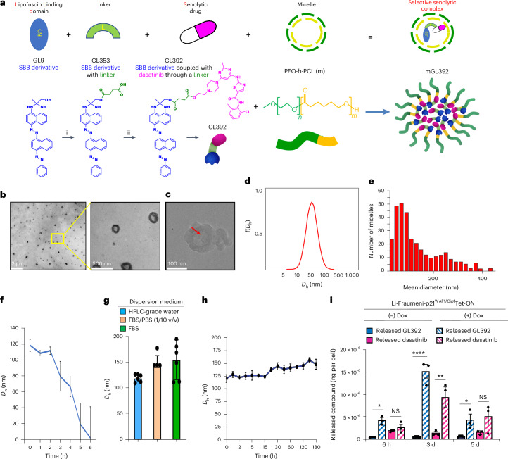
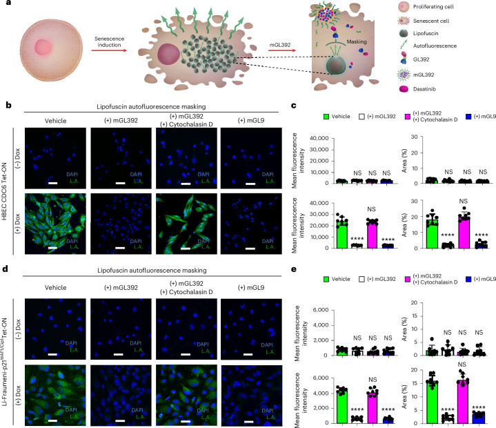
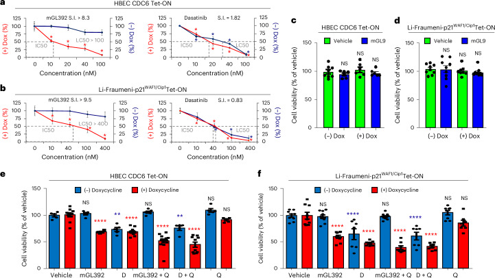
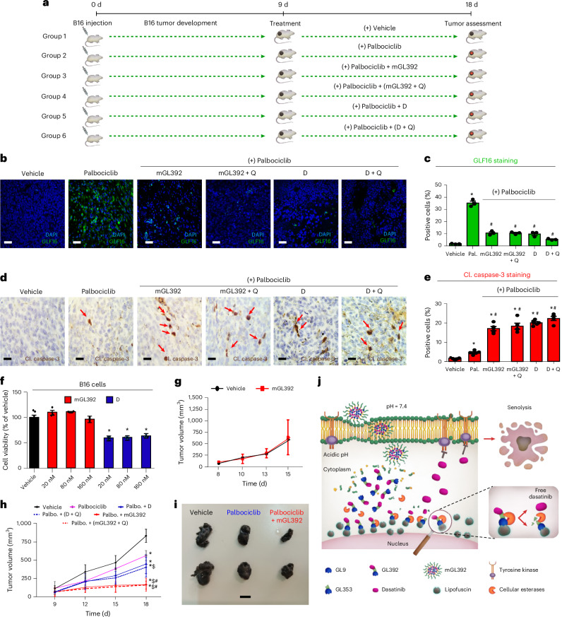
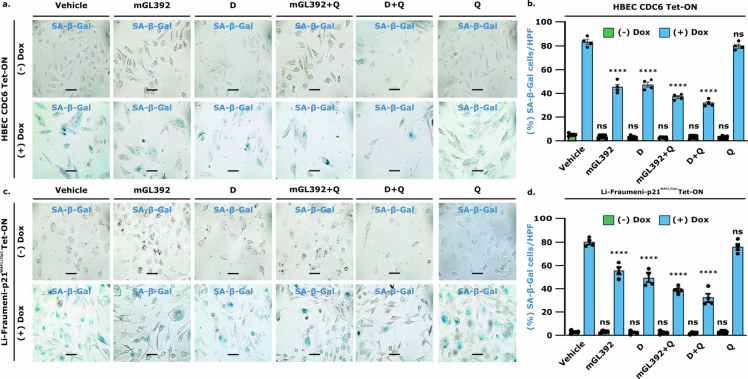
The emerging field of senolytics is centered on eliminating senescent cells to block their contribution to the progression of age-related diseases, including cancer, and to facilitate healthy aging. Enhancing the selectivity of senolytic treatments toward senescent cells stands to reduce the adverse effects associated with existing senolytic interventions. Taking advantage of lipofuscin accumulation in senescent cells, we describe here the development of a highly efficient senolytic platform consisting of a lipofuscin-binding domain scaffold, which can be conjugated with a senolytic drug via an ester bond. As a proof of concept, we present the generation of GL392, a senolytic compound that carries a dasatinib senolytic moiety. Encapsulation of the GL392 compound in a micelle nanocarrier (termed mGL392) allows for both in vitro and in vivo (in mice) selective elimination of senescent cells via targeted release of the senolytic agent with minimal systemic toxicity. Our findings suggest that this platform could be used to enhance targeting of senotherapeutics toward senescent cells.


Conflict of interest statement

Competing interests: Described compounds are in patent pending status: (1) Greek Patent Application No. 20240100309, (2) UK Patent Application No. GB2406749.8 and (3) international Patent Cooperation Treaty filing. Patent competing interests concern N.K. and V.G.G. only. The other authors declare no competing interests.

Figures

Fig. 1
Fig. 1. The mGL392 compound constitutes a micellated dasatinib-conjugated LBD scaffold for effective targeting of senescent cells.
a, Upper panel: the GL392 building platform consists of three regions: the LBD, the senolytic compound and an esteric linker connecting the LBD to the senolytic compound. In the case of GL392, dasatinib was selected as a potent senolytic drug. Lower panel: GL9 contains the LBD and is coupled with dasatinib (purple) through a succinic linker (green) to generate GL392. For more effective dasatinib delivery and specificity, GL392 is encapsulated in PEO-b-PCL micelles to create the senolytic mGL392 compound. bh, Physicochemical characterization of mGL392. Structure and physicochemical properties of micelle-GL392 were verified using TEM (the yellow inset is magnified on the right frame) (b) and cryo-TEM (red arrow indicates the electron-lucent core of mGL392 where GL392 is successfully incorporated) (c). Homogeneity of the micelle preparation was verified by evaluation of mGL392 particle distribution based on size (d) using DLS and number (e) using TEM. Efficient degradation of mGL392 at acidic pH (4.5) was determined by measuring the mean Dh of the particles during a 6-h timecourse using DLS (n = 3 independent chemical experiments) (f). Stability of the complex in cell culture relevant media was verified at 37 °C (n = 5 independent chemical experiments) (g) and during the course of a 180-h timeframe (n = 5 independent chemical experiments) (h) using DLS. i, Released GL392 or dasatinib was traced in lysates of senescent (+Dox) or non-senescent (−Dox) Li-Fraumeni-p21WAF1/Cip1 Tet-ON cells with LC–MS, after incubation with mGL392 for the indicated time (n = 3 biological replicates). * P: 0.01–0.05, ** P: 0.001–0.01, **** P < 0.0001, two-way ANOVA (i). Error bars indicate s.d. NS, non-significant. Data are presented as mean values ± s.d. from at least three independent biological replicates (fi).
Fig. 2
Fig. 2. The mGL392 compound selectively accumulates within senescent cells.
a, Proper internalization of mGL392 and specific binding of its cargo to lipofuscin was validated based on lipofuscin auto-fluorescence. During senescence, cells accumulate lipofuscin that emits auto-fluorescence. Upon binding to lipofuscin, mGL392 masks emitted auto-fluorescence signals. b, Representative images of senescent (+Dox) or non-senescent (−Dox) HBEC CDC6 Tet-ON, treated or not with cytochalasin D (an actin polymerization inhibitor) and mGL392/mGL9 (10 nM) and evaluated for lipofuscin auto-fluorescence. c, Mean fluorescence intensity and percent area of lipofuscin auto-fluorescence in b were quantified using ImageJ (n = 8 biological replicates). d, Representative images of senescent (+Dox) or non-senescent (−Dox) Li-Fraumeni-p21WAF1/Cip1 Tet-ON cells, treated or not with cytochalasin D (an actin polymerization inhibitor) and mGL392/mGL9 (10 nM) and evaluated for lipofuscin auto-fluorescence. e, Mean fluorescence intensity and percent area of lipofuscin auto-fluorescence in d were quantified using ImageJ (n = 8 biological replicates). Objective: ×40. Scale bar, 25 μm. **** P < 0.0001, one-way ANOVA (statistical denotations represent comparisons to vehicle (PBS)). Error bars indicate s.d. NS, non-significant. Data are presented as mean values ± s.d. from eight independent biological replicates. L.A., lipofuscin autofluorescence.
Fig. 3
Fig. 3. Selectivity of mGL392-mediated senolysis is superior to that mediated by free dasatinib.
a,b, Determination of the selectivity index (SI) for mGL392 compared to free dasatinib in the two inducible cellular senescence systems in HBEC CDC6 Tet-ON (a) and Li-Fraumeni-p21WAF1/Cip1 Tet-ON (b) cells. In both cases, cells were treated with Dox for senescence entry and subsequently with incremental concentrations of mGL392 (0–400 nM) or free dasatinib (0–400 nM). IC50 confers 50% reduction of senescent cells (red line) (senolytic effect), whereas LC50 eliminates 50% of non-senescent cells (blue line) (cytotoxic effect). SI was calculated as the LC50/IC50 ratio. Data expressed are as percent mean ± s.e.m. of vehicle (PBS), n = 9 (biological replicates); blue and red stars represent statistical significance compared to respective (−) and (+) Dox vehicles, * P < 0.05 compared to vehicle, one-way ANOVA test. c,d, GL9 LBD does not exert senolytic or cytotoxic actions. GL9 was encapsulated in a micelle complex and was applied in HBEC CDC6 Tet-ON (c) and Li-Fraumeni-p21WAF1/Cip1 Tet-ON (d) cells. Cell viability was determined by MTT assay. Data are expressed as percent mean ± s.d. of total cells, n = 9 (biological replicates); NS, non-significant compared to respective vehicle (two-sided t-test). e,f, Validation of the senolytic properties of mGL392 in the HBEC CDC6 Tet-ON (e) and Li-Fraumeni-p21WAF1/Cip1 Tet-ON (f) cellular systems. Senescent and non-senescent cells were treated with vehicle (PBS), mGL392 (10 nM in the case of HBEC CDC6 Tet-ON and 20 nM in case of Li-Fraumeni-p21WAF1/Cip1 Tet-ON cells) or mGL392+quercetin (mGL392+Q, 10 nM or 20 nM and 10 μΜ, respectively). Free dasatinib (D, 10 nM and 20 nM for HBEC CDC6 Tet-ON and Li-Fraumeni-p21WAF1/Cip1 Tet-ON cells, respectively), quercetin (Q, 10 μΜ) and free dasatinib+quercetin (D+Q) were used as reference. Cell viability was assessed by MTT assay, 4–6 d after treatment. Results are expressed as mean percent of vehicle ± s.d. Blue and red stars in each condition represent statistical significance compared to (−) and (+) Dox vehicles, respectively. ** P: 0.001–0.01, **** P < 0.0001, two-way ANOVA test (e and f). Error bars indicate s.d. Data are presented as mean values ± s.d, n = 14 (biological replicates).
Fig. 4
Fig. 4. mGL392 selectively eliminates senescent cells in vitro in HBEC CDC6 Tet-ON cells.
a, Selectivity of mGL392 senolysis was verified by visualization of apoptotic (Cl. Caspase-3+, red) and senescent (GLF16+, green) cells in senescent (+Dox) or non-senescent (−Dox) HBEC CDC6 Tet-ON cells. b, Quantification of immunofluorescence in c. Blue, green and red stars in each condition represent statistical significance compared to respective (−) and (+) Dox vehicles. Results are expressed as percent positive (senescent or apoptotic) cells counted against DAPI-stained nuclei inspected from at least 10 optical fields per sample (n = 3 biological replicates). D, dasatinib; Q, quercetin. Objective: ×40. Scale bar, 25 μm. Blue, green and red stars in each condition represent statistical significance compared to respective (−) and (+) Dox vehicles. Insets represent digital magnifications of representative (indicated) cells within the presented frame. **** P < 0.0001, two-way ANOVA test. Error bars indicate s.d. Data are presented as mean values ± s.d. from three independent biological replicates. NS, non-significant.
Fig. 5
Fig. 5. mGL392 selectively eliminates senescent cells in vitro in Li-Fraumeni-p21WAF1/Cip1 Tet-ON cells.
a, Selectivity of mGL392 senolysis was verified by visualization of apoptotic (Cl. Caspase-3+, red) and senescent (GLF16+, green) cells in senescent (+Dox) or non-senescent (−Dox) Li-Fraumeni-p21WAF1/Cip1 Tet-ON cells. b, Quantification of immunofluorescence in c. Blue, green and red stars in each condition represent statistical significance compared to respective (−) and (+) Dox vehicles. Results are expressed as percent positive (senescent or apoptotic) cells counted against DAPI-stained nuclei inspected from at least 10 optical fields per sample (n = 3 biological replicates). D, dasatinib; Q, quercetin. Objective: ×40. Scale bar, 25 μm. Blue, green and red stars in each condition represent statistical significance compared to respective (−) and (+) Dox vehicles. Insets represent digital magnifications of representative (indicated) cells within the presented frame. **** P < 0.0001, two-way ANOVA test. Error bars indicate s.d. Data are presented as mean values ± s.d. from three independent biological replicates. NS, non-significant.
Fig. 6
Fig. 6. mGL392 induces targeted senolysis in 3D patient-derived lung organoids.
a, Overview of the experimental workflow. AOs were generated from surgically resected healthy lung tissue. Organoid cultures were treated with H2O2 for senescence induction or left untreated. In both cases, the organoid medium was supplemented with vehicle (PBS), mGL392 (500 nM), dasatinib (D, 500 nM), quercetin (Q, 10 μΜ), dasatinib and quercetin (D+Q) or mGL392 and quercetin (mGL392+Q) for 4 d. Selective senolysis was evaluated by cell viability measurements and quantification of senescent and apoptotic cells by immunofluorescence. b, Organoid cell viability was determined using a proteasome activity assay. Results are presented as percent of vehicle. Blue and red stars in each condition represent statistical significance compared to (−) and (+) H2O2 vehicles, respectively (n = 3 biological replicates). c,d, Apoptosis (Cl. Caspase-3 immunoreactivity, red) and senescence (GLF16 staining, green) assessment was performed to verify the selective senolytic activity of mGL392. c, Representative images of confocal microscopy. Objective: ×20. Scale bar, 10 μm. d, Quantification of immunofluorescence analysis in c. Results are expressed as percent positive (senescent or apoptotic) cells counted against DAPI-stained nuclei inspected from at least 10 optical fields per sample (n = 3 biological replicates). Blue, red and green stars in each condition represent statistical significance compared to respective (−) and (+) H2O2 vehicles. * P: 0.001–0.05, ** P: 0.001–0.01, **** P < 0.0001, two-way ANOVA test. Error bars indicate s.d. Data are presented as mean values ± s.d. from three independent biological replicates. NS, non-significant.
Fig. 7
Fig. 7. mGL392 enables efficient delivery and selective release of the senolytic agent in vivo.
a, The in vivo mGL392 senolytic potential was challenged in a murine model of palbociblib-induced senescence. Timeline of in vivo experiment. Murine melanomas developed upon subcutaneous injection of B16 cells. Mice were split into six groups and received vehicle (normal saline), palbociclib (Pal.), palbociclib/mGL392 (mGL392), palbociclib/mGL392/quercetin (mGL392 + Q), palbociclib/dasatinib (D) and palbociclib/dasatinib/quercetin (D + Q) daily for 9 d. Palbociclib was administered at 2.5 mg per mouse, mGL392 at 0.015 mg per mouse, dasatinib at 0.125 mg per mouse and quercetin at 1.25 mg per mouse. b,c, Representative immunofluorescence images displaying senescent cells (GLF16+, green) (b) and quantification of senescent cell populations in the tumors (c). Results are expressed as percent positive senescent cells counted against DAPI-stained nuclei from at least 10 inspected optical fields per sample. Data are presented as mean values ± s.d. of total cells, n = 5 (biological replicates), * P < 0.05 compared to vehicle, # P < 0.05 compared to the palbociclib group by one-way ANOVA. Objective: ×20. Scale bar, 10 μm. d,e, Effective mGL392-mediated senolysis leads to enhanced tumor cell apoptosis. Representative images of Cl. Caspase-3 staining (cells depicted by red arrows) (d) and quantification of analysis (e). Results are expressed as percent mean ± s.d. of total cells, n = 5 (biological replicates), * P < 0.05 compared to vehicle, #P < 0.05 compared to the palbociclib group by one-way ANOVA. Data are presented as mean values ± s.d. Objective: ×20. Scale bar, 10 μm. f,g, mGL392 does not affect non-senescent B16 melanoma growth in vitro (n = 4 biological replicates) (f) or in vivo (n = 6 biological replicates) (g), as assessed by MTT cell viability and tumor size measurements, respectively. Results are expressed as mean ± s.d., * P < 0.05 compared to vehicle by one-way ANOVA. Data expressed are presented as mean values ± s.d. h, mGL392 enhances the melanoma-reducing action of palbociclib. Tumor sizes were measured every 3 d. Data are presented as mean values ± s.e.m., n = 8 (biological replicates), * P < 0.05 compared to vehicle, #P < 0.05 compared to dasatinib, $P < 0.05 compared to palbociclib by one-way ANOVA. i, Representative tumors from h. Scale bar, 5 mm. j, Proposed model of mGL392 action. Palbo., palbociclib.
Extended Data Fig. 1
Extended Data Fig. 1. mGL392 selectively delivers GL392 in senescent cells followed by Dasatinib release.
(a) Schematic representation of the dynamic process of Dasatinib release. mGL392 preferably enters senescent cells delivering its GL392 cargo. Dasatinib release initiates 6 h upon mGL392 treatment and peaks 3 days later. (b) Representative output of LC-MS analysis of senescent (+Dox) versus non- senescent (-Dox) Li-Fraumeni-p21WAF1/Cip1 Tet-ON cell lysates from cells treated with mGL392 for 3 days. Peaks depict relative abundance of the released GL392 and the released Dasatinib traced in the cell lysates. Peaks were identified based on prepared GL392 and Dasatinib stock solutions. Lapatinib served as an internal standard of the analysis.
Extended Data Fig. 2
Extended Data Fig. 2. Verification of mGL392-induced senolysis by SA-β-gal staining.
HBEC CDC6 Tet-ON (a, b) and Li-Fraumeni-p21WAF1/Cip1 Tet-ON (c, d) cells entered senescence upon Doxycycline (Dox) treatment. Senescent and respective non-senescent counterparts were subsequently treated with Vehicle (PBS), mGL392 (10 nM in the case of HBEC CDC6 Tet-ON and 20 nM in the case of Li-Fraumeni-p21WAF1/Cip1 Tet-ON cells) or mGL392+Quercetin (mGL392+Q, 10 or 20 nM and 10 μΜ, respectively), free Dasatinib (D, 10 and 20 nM for HBEC CDC6 Tet-ON and Li-Fraumeni-p21WAF1/Cip1 Tet-ON cells, respectively), Quercetin (Q, 10 μΜ) and free Dasatinib+Quercetin (D + Q) for 4-6 days and stained for SA-β-gal. (a, c) Representative images. Objective 20x. Scale bar: 10 μm. (b, d) Quantification of SA-β-gal(+) cells, n = 4 (biological replicates), **** p < 0.0001 compared to respective vehicle, two-way ANOVA test. Error bars indicate SD. Data expressed are presented as mean values ± SEM.
Extended Data Fig. 3
Extended Data Fig. 3. Verification of mGL392-induced senolytic activity via flow cytometry-based apoptotic assessment.
HBEC CDC6 Tet-ON (a, c) and Li-Fraumeni-p21WAF1/Cip1 Tet-ON (b, d) cells were treated with Doxycycline (Dox) to induce senescence. Senescent cells and their non-senescent counterparts were treated with Vehicle (PBS), mGL392 (10 nM in the case of HBEC CDC6 Tet-ON and 20 nM in the case of Li-Fraumeni-p21WAF1/Cip1 Tet-ON cells) or mGL392+Quercetin (mGL392+Q, 10 or 20 nM and 10 μΜ, respectively), free Dasatinib (D, 10 and 20 nM for HBEC CDC6 Tet-ON and Li-Fraumeni-p21WAF1/Cip1 Tet-ON cells, respectively), Dasatinib+Quercetin (D + Q, 10 or 20 nM and 10 μΜ, respectively) and Quercetin (Q, 10 μΜ) for 4-6 days and subsequently stained with Annexin V-FITC and PI for evaluation of apoptotic and dead cells, respectively. (a, b) Representative density plots from flow cytometry analysis. (c, d) Quantification of flow cytometry results presented as mean ± SEM, n = 4 (biological replicates), **** p < 0.0001; n.s. non-significant compared to respective vehicle by two-way ANOVA. Data expressed are presented as mean values ± SEM.
Extended Data Fig. 4
Extended Data Fig. 4. Lung organoid stainings directly comparing mGL392 treatment with free agents.
Individual staining panels of organoids from Fig. 6c. From left to right, representative split-channel image overlays of DAPI (blue), GLF16 (green) and Cl. Caspase-3 (red) staining, and respective composites. Objective 20x. Scale bar: 10 μm. Data shown are presented as mean values ± SD from 3 independent biological replicates.
Extended Data Fig. 5
Extended Data Fig. 5. In vivo administration of mGL392 effectively eliminates SA-β-gal(+) cells in the tumor without conferring cytotoxicity.
(a) Representative pictures of excised mouse tumors stained for SA-β-gal and (b) quantification of SA-β-gal(+) cells in (a) (n = 4, biological replicates). (c) Representative tumors from Fig. 7i, Scale bar: 5 mm. (d) Sera of vehicle (normal saline), mGL392 and Dasatinib-treated mice were analyzed for common toxicology markers associated with vital organ damage. Results are expressed as mean ± SD (n = 9 biological replicates). CREA: Creatinine; ALB: Albumin; TP: Total Protein; TBIL: Total Bilirubin; ALP: Alkaline Phosphatase; γ-GT: γ-glutamyltransferase; ALT: Alanine Transaminase; AST: Aspartate Aminotransferase; BUN: Blood Urea Nitrogen; CPK: Creatine Phosphokinase. (e) Vital organs (lung, liver, kidney, heart, spleen) and hindlimb muscle samples of all mice were excised and stained with hematoxylin and eosin for histopathological evaluation. (f) Monocyte populations in the blood of mice receiving vehicle (normal saline) or mGL392 were quantified using flow cytometry and found unaffected, n = 4 (biological replicates). Data are presented as mean values +/−SEM, *p < 0.05 compared to vehicle, #p < 0.05 compared to Palbociclib. n.s.; non-significant. Two-way ANOVA test (b), one-way ANOVA test (d), two-sided t-test (f) were used. Objectives 10x, 20x. Scale bars: 50 μm and 10 μm.

Similar articles

Cited by

References

    1. Gorgoulis, V. et al. Cellular senescence: defining a path forward. Cell31, 813–827 (2019). - PubMed
    1. Ogrodnik, M. et al. Guidelines for minimal information on cellular senescence experimentation in vivo. Cell187, 4150–4175 (2024). - PMC - PubMed
    1. Zampetidis, C. P. et al. A recurrent chromosomal inversion suffices for driving escape from oncogene-induced senescence via subTAD reorganization. Mol. Cell81, 4907–4923 (2021). - PubMed
    1. Bartkova, J. et al. Oncogene-induced senescence is part of the tumorigenesis barrier imposed by DNA damage checkpoints. Nature444, 633–637 (2006). - PubMed
    1. Halazonetis, T. D., Gorgoulis, V. G. & Bartek, J. An oncogene-induced DNA damage model for cancer development. Science319, 1352–1355 (2008). - PubMed

LinkOut - more resources