

Therapeutic role of aripiprazole in cartilage defects explored through a drug repurposing approach
- PMID: 39730885
- PMCID: PMC11680846
- DOI: 10.1038/s41598-024-82177-1
Therapeutic role of aripiprazole in cartilage defects explored through a drug repurposing approach
Abstract
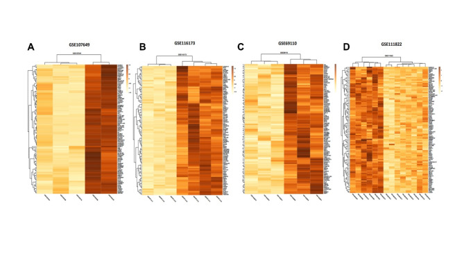
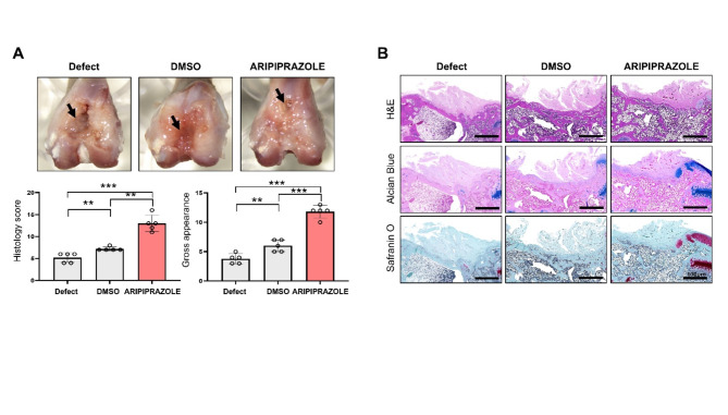
Articular cartilage has a limited regenerative capacity, resulting in poor spontaneous healing of damaged tissue. Despite various scientific efforts to enhance cartilage repair, no single method has yielded satisfactory results. With rising drug development costs, drug repositioning has emerged as a viable alternative. This study aimed to identify a drug capable of improving cartilage defects by analyzing chondrogenesis-related microarray data from the Gene Expression Omnibus (GEO) public database. We utilized datasets GSE69110, GSE107649, GSE111822, and GSE116173 to identify genes associated with cartilage differentiation, employing StringTie for differential gene expression analysis and extracting drug data from the Drug-Gene Interaction database. Additionally, we aimed to verify the cartilage regeneration potential of the identified drug through experiments using cellular and animal models. We evaluated the effects of aripiprazole on adipose-derived mesenchymal stem cells (ADMSCs) and chondrocytes using qRT-PCR and a 3D pellet culture system. In vivo, we assessed cartilage restoration by combining aripiprazole with a scaffold and implanting it into artificially induced cartilage defects in Sprague-Dawley rats. Subsequent mRNA sequencing provided insights into the mechanistic pathways involved. Our results showed that aripiprazole significantly increased mRNA expression of COL2A1 and SOX9, markers of chondrogenesis, and promoted chondrogenic condensation in vitro. Furthermore, aripiprazole effectively enhanced cartilage regeneration in the rat model. KEGG pathway and Gene Ontology Biological Processes (GOBP) analyses of the mRNA sequencing data revealed that aripiprazole upregulated genes related to ribosomes and cytoplasmic translation, thereby facilitating chondrogenesis. In conclusion, our findings suggest that aripiprazole is a promising candidate for improving damaged cartilage, offering a novel approach to cartilage regeneration.
© 2024. The Author(s).
Conflict of interest statement
Declarations. Competing interests: The authors declare no competing interests.
Figures


Similar articles
-
Cladophora glomerata methanolic extract promotes chondrogenic gene expression and cartilage phenotype differentiation in equine adipose-derived mesenchymal stromal stem cells affected by metabolic syndrome.Stem Cell Res Ther. 2019 Dec 17;10(1):392. doi: 10.1186/s13287-019-1499-z. Stem Cell Res Ther. 2019. PMID: 31847882 Free PMC article.
-
Oral Administration of Strontium Gluconate Effectively Reduces Articular Cartilage Degeneration Through Enhanced Anabolic Activity of Chondrocytes and Chondrogenetic Differentiation of Mesenchymal Stromal Cells.Biol Trace Elem Res. 2020 Feb;193(2):422-433. doi: 10.1007/s12011-019-01711-9. Epub 2019 May 3. Biol Trace Elem Res. 2020. PMID: 31054068
-
Rapamycin-Induced Autophagy Promotes the Chondrogenic Differentiation of Synovium-Derived Mesenchymal Stem Cells in the Temporomandibular Joint in Response to IL-1β.Biomed Res Int. 2020 Oct 22;2020:4035306. doi: 10.1155/2020/4035306. eCollection 2020. Biomed Res Int. 2020. PMID: 33145347 Free PMC article.
-
Mechanotransduction and Stiffness-Sensing: Mechanisms and Opportunities to Control Multiple Molecular Aspects of Cell Phenotype as a Design Cornerstone of Cell-Instructive Biomaterials for Articular Cartilage Repair.Int J Mol Sci. 2020 Jul 29;21(15):5399. doi: 10.3390/ijms21155399. Int J Mol Sci. 2020. PMID: 32751354 Free PMC article. Review.
-
Should we use cells, biomaterials, or tissue engineering for cartilage regeneration?Stem Cell Res Ther. 2016 Apr 18;7(1):56. doi: 10.1186/s13287-016-0314-3. Stem Cell Res Ther. 2016. PMID: 27089917 Free PMC article. Review.
References
-
- Charlier, E. et al. Chondrocyte dedifferentiation and osteoarthritis (OA). Biochem. Pharmacol.165, 49–65 (2019). - PubMed
-
- Johnson, V. L. & Hunter, D. J. The epidemiology of osteoarthritis. Best Pract. Res. Clin. Rheumatol.28, 5–15 (2014). - PubMed
-
- Zelinka, A., Roelofs, A. J., Kandel, R. A. & De Bari, C. Cellular therapy and tissue engineering for cartilage repair. Osteoarthr. Cartil.30, 1547–1560 (2022). - PubMed
MeSH terms
Substances
Grants and funding
- RS-2023-00210067 and 2022R1A2C2005916/National Research Foundation of Korea
- RS-2023-00210067 and 2022R1A2C2005916/National Research Foundation of Korea
- RS-2023-00210067 and 2022R1A2C2005916/National Research Foundation of Korea
- RS-2023-00210067 and 2022R1A2C2005916/National Research Foundation of Korea
- RS-2023-00210067 and 2022R1A2C2005916/National Research Foundation of Korea
LinkOut - more resources
Full Text Sources
Molecular Biology Databases
Research Materials
