

Prediction of grading of ovarian endometrioid carcinoma using conventional MRI features
- PMID: 39730935
- PMCID: PMC12052869
- DOI: 10.1007/s11604-024-01727-9
Prediction of grading of ovarian endometrioid carcinoma using conventional MRI features
Abstract
Objective: The purpose of this study was to evaluate MRI findings of ovarian endometrioid carcinoma (OEC) as a predictor of histological grade.
Materials and methods: This study included 60 patients with histopathologically confirmed OEC (20, 30, and 10 with grades 1, 2, and 3, respectively). Clinical and MRI results were retrospectively reviewed. We compared the following parameters between the three grades: age, tumor markers, presence of uterine corpus cancer, bilaterality, configuration, peritoneal dissemination, abnormal ascites, signal intensities of cystic and solid components, tumor size, and apparent diffusion coefficient (ADC) values of solid components.
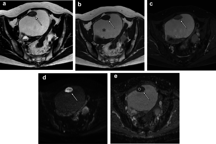
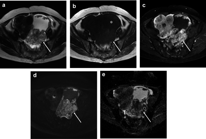
Results: T1-hyperintense cysts were more common in grade 1 than in grades 2-3 OEC (80% vs. 60%, vs. 40%, p < 0.05). The signal intensity ratio between the cystic components with the largest solid component and muscle (1.49 vs. 1.08 vs. 0.98, p < 0.05) was higher in grade 1 than in grades 2-3 OEC. Necrosis within solid components was less common in grade 1 than in grades 2-3 OEC (31% vs. 68% vs. 88%, p < 0.05), and the ADC values of solid components were higher in grade 1 than in grades 2-3 OEC (1.10 vs. 0.99 vs. 0.79 × 10⁻³ mm2/sec, p < 0.05). There were no significant differences in other factors.
Conclusion: On T1-weighted images, grade 1 OEC showed a higher signal intensity in the cystic components than grades 2-3 OEC. Necrosis and lower ADC values were more frequently observed in grades 2-3 than in grade 1 OEC.
Keywords: Carcinoma; Endometrioid; MRI; Ovary; Tumor grading.
© 2024. The Author(s).
Conflict of interest statement
Declarations. Conflict of interest: The authors declare that they have no conflict of interest. Data availability: Data will be made available on reasonable request.
Figures
Similar articles
-
Primary ovarian endometrioid adenocarcinoma: magnetic resonance imaging findings including a preliminary observation on diffusion-weighted imaging.J Comput Assist Tomogr. 2015 May-Jun;39(3):401-5. doi: 10.1097/RCT.0000000000000210. J Comput Assist Tomogr. 2015. PMID: 25978592
-
Endometrioid adenocarcinoma of the ovary: MRI findings with emphasis on diffusion-weighted imaging for the differentiation of ovarian tumors.Acta Radiol. 2016 Jun;57(6):758-66. doi: 10.1177/0284185115599805. Epub 2015 Aug 24. Acta Radiol. 2016. PMID: 26307063
-
Morphological and Immunohistochemical Reevaluation of Tumors Initially Diagnosed as Ovarian Endometrioid Carcinoma With Emphasis on High-grade Tumors.Am J Surg Pathol. 2016 Mar;40(3):302-12. doi: 10.1097/PAS.0000000000000550. Am J Surg Pathol. 2016. PMID: 26551621 Free PMC article.
-
MRI for differentiating ovarian endometrioid adenocarcinoma from high-grade serous adenocarcinoma.J Ovarian Res. 2015 Apr 30;8:26. doi: 10.1186/s13048-015-0154-2. J Ovarian Res. 2015. PMID: 25926038 Free PMC article.
-
TCGA molecular subgroups of endometrial carcinoma in ovarian endometrioid carcinoma: A quantitative systematic review.Gynecol Oncol. 2021 Nov;163(2):427-432. doi: 10.1016/j.ygyno.2021.08.011. Epub 2021 Aug 24. Gynecol Oncol. 2021. PMID: 34446267
References
-
- Köbel M, Huntsman D, Lim D, McCluggage W, Rabban J, Shih I. Endometrioid carcinoma of the ovary. WHO classification of Female Genital Tumours. 5th ed. Lyon, France: IARC Press; 2020. p. 58–61.
-
- Swift BE, Covens A, Mintsopoulos V, Parra-Herran C, Bernardini MQ, Nofech-Mozes S, et al. The effect of complete surgical staging and adjuvant chemotherapy on survival in stage I, grade 1 and 2 endometrioid ovarian carcinoma. Int J Gynecol Cancer. 2022. 10.1136/ijgc-2021-003112. - PubMed
-
- Assem H, Rambau PF, Lee S, Ogilvie T, Sienko A, Kelemen LE, et al. High-grade endometrioid carcinoma of the ovary: a clinicopathologic study of 30 cases. Am J Surg Pathol. 2018. 10.1097/pas.0000000000001016. - PubMed
-
- Berek JS, Matias-Guiu X, Creutzberg C, Fotopoulou C, Gaffney D, Kehoe S, et al. FIGO staging of endometrial cancer: 2023. Int J Gynaecol Obstet. 2023. 10.1002/ijgo.14923. - PubMed
-
- Network NCC. Ovarian cancer including fallopian tube cancer and primary peritoneal cancer (version 3.2024). 2024(August 22, 2024).
MeSH terms
LinkOut - more resources
Full Text Sources
Medical
