. 2024 Dec 28;14(1):31112.
doi: 10.1038/s41598-024-82325-7.

Therapy response monitoring in blood plasma from esophageal adenocarcinoma patients using cell-free DNA methylation profiling

Affiliations

Therapy response monitoring in blood plasma from esophageal adenocarcinoma patients using cell-free DNA methylation profiling

Kathleen Schoofs et al. Sci Rep. .

Abstract

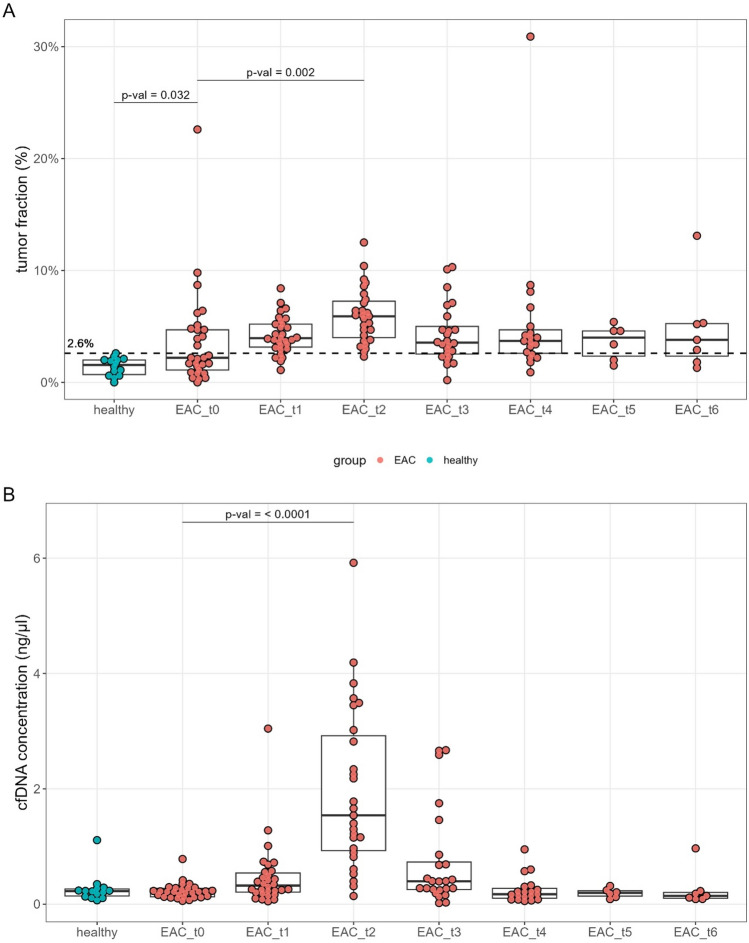
Esophageal adenocarcinoma (EAC) is an aggressive cancer characterized by a high risk of relapse post-surgery. Current follow-up methods (serum carcinoembryonic antigen detection and PET-CT) lack sensitivity and reliability, necessitating a novel approach. Analyzing cell-free DNA (cfDNA) from blood plasma emerges as a promising avenue. This study aims to evaluate the cost-effective and genome-wide cell-free reduced representation bisulfite sequencing (cfRRBS) method combined with computational deconvolution for effective disease monitoring in EAC patients. cfDNA methylation profiling with cfRRBS was performed on 162 blood plasma samples from 33 EAC cancer patients and 28 blood plasma samples from 20 healthy donors. The estimated tumor fraction for EAC patients at the time of diagnosis was significantly different from the healthy donor plasma samples (one-sided Wilcoxon rank-sum test: p-value = 0.032). Tumor fractions above 15% and focal gains/amplifications in MYC (chr8), KRAS (chr12), EGFR (chr7) and NOTCH2 (chr1) were observed in four samples of distinct patients at the time metastatic disease was detected. This study showed feasibility to estimate tumor fractions in blood plasma of EAC patients based on cfDNA methylation using cfRRBS and computational deconvolution. Nevertheless, in this study only cancer patients with evidence of metastatic disease show high tumor fractions and copy number alterations.

Keywords: Blood plasma; DNA methylation; Esophageal adenocarcinoma; Liquid biopsy; cfDNA.


Conflict of interest statement

Declarations. Competing interests: The authors declare no competing interests. Ethics: Written informed consent was obtained from all cancer patients and healthy donors. Sample collection was approved by the ethics committee of Ghent University Hospital (registration numbers B670201628317, B670201628319 and B670201733701). The research was conducted according to the local legislation and institutional requirements.

Figures

Fig. 1
Fig. 1
Dynamic change of tumor fractions and cfDNA concentration in plasma from EAC patients. (A) Boxplots of estimated EAC tumor fractions in the patient cfDNA sampled at different timepoints and in the 14 healthy donor samples. Based on the highest estimated tumor fraction of the healthy donor samples, a cut-off was set at 2.6%. (B) cfDNA concentration (ng/µL) in blood plasma of healthy donors and EAC patients at the different timepoints.
Fig. 2
Fig. 2
Tumor fractions in matched plasma samples collected in two blood collection tubes. No significant difference (paired Wilcoxon rank-sum test, p-value = 0.46) in EAC tumor fraction between six matched healthy donor plasma samples collected in citrate and EDTA blood collection tubes.
Fig. 3
Fig. 3
Dynamic change in estimated tumor fraction in longitudinally collected blood plasma samples from 33 EAC patients. (A) Heatmap showing estimated tumor fractions per patient and sampled timepoint. The x-axis indicates the time in months relative to the time of surgery (timepoint t1). Grey boxes indicate tumor fractions below the cut-off of 2.6%, blue between 2.6% and 15% and dark blue more than 15%. (BD) Timelines showing increasing tumor fractions of three patients that eventually presented with a tumor fraction that is higher than 15% (after surgery).
Fig. 4
Fig. 4
CNV profiles based on cfRRBS data show aberrations in plasma samples with tumor fractions above 15%. (A) CNV profile of ID25_EAC_4, showing a focal gain/amplification in chromosome 8; (B) CNV profile of ID48_EAC_4, showing a focal gain/amplification in chromosome 12; (C) CNV profile of ID55_EAC_3, showing a focal gain/amplification in chromosome 7; (D) complete CNV profile of ID58_EAC_0 and a zoomed in profile of chromosome 1. The profile of this patient blood sample collected at the time of diagnosis showed a focal gain/amplification in chromosome 1.

Similar articles

References

    1. Oppedijk, V. et al. Patterns of recurrence after surgery alone versus preoperative chemoradiotherapy and surgery in the CROSS trials. J. Clin. Oncol.32, 385–391. 10.1200/jco.2013.51.2186 (2014). - PubMed
    1. Bruzzi, J. F. et al. PET/CT of esophageal cancer: its role in clinical management. Radiographics27, 1635–1652. 10.1148/rg.276065742 (2007). - PubMed
    1. Roedl, J. B. et al. Assessment of treatment response and recurrence in esophageal carcinoma based on tumor length and standardized uptake value on positron emission tomography-computed tomography. Ann. Thorac. Surg.86, 1131–1138. 10.1016/j.athoracsur.2008.05.019 (2008). - PubMed
    1. Valkema, M. J. et al. Accuracy of (18)F-FDG PET/CT in predicting residual disease after neoadjuvant chemoradiotherapy for esophageal cancer. J. Nucl. Med.60, 1553–1559. 10.2967/jnumed.118.224196 (2019). - PubMed
    1. Moyes, L. H., Anderson, J. E. & Forshaw, M. J. Proposed follow up programme after curative resection for lower third oesophageal cancer. World J. Surg. Oncol.8, 75. 10.1186/1477-7819-8-75 (2010). - PMC - PubMed

Supplementary concepts