

Early response of anti-vascular endothelial growth factor (anti-VEGF) in diabetic macular edema (DME) management: microperimetry and optical coherence tomography (OCT) findings: a pilot study at national eye center of third world country
- PMID: 39731082
- PMCID: PMC11674592
- DOI: 10.1186/s12886-024-03744-8
Early response of anti-vascular endothelial growth factor (anti-VEGF) in diabetic macular edema (DME) management: microperimetry and optical coherence tomography (OCT) findings: a pilot study at national eye center of third world country
Abstract
Purpose: To evaluate early response of retinal sensitivity (RS) and retinal morphology in diabetic macular edema (DME) patients after intravitreal anti-vascular endothelial growth factor (anti-VEGF) treatment.
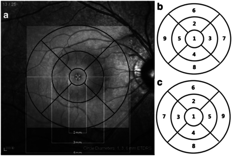
Methods: Sixteen eyes of 12 DME patients were included in this study conducted prospectively. All eyes underwent functional and morphologic examination of the macular area using microperimetry and optical coherence tomography (OCT) before and after intravitreal anti-VEGF injection. To determine significant differences between the values, paired t test was used. A correlation between CMT and RS was made using Spearman's test.
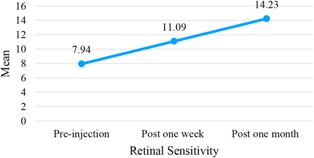
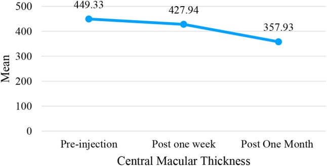
Results: Patients were evaluated at baseline, one week and one month after injection. The central macular thickness (CMT) decreased significantly from 449.33 ± 100.79 μm to 427.94 ± 85.76 μm to 357.93 ± 75.92 μm. The RS improved significantly from 7.94 ± 6.43 dB to 11.09 ± 7.42 dB at one week and to 14.22 ± 7.66 dB at one month after treatment. The CMT was significant negatively correlated to RS (r=-0.259, p = < 0.001), with decay of 0.025 dB for every 1 μm increase of CMT.
Conclusions: Retinal thickening due to DME can be adequately quantified using OCT, while microperimetry can offer information about retinal sensitivity in the exact location. Therefore, microperimetry can be a useful tool in predicting the functional outcome and determining the efficacy of anti-VEGF treatment for DME patients.
Keywords: Anti-VEGF response; Diabetic macular edema; Macular thickness; Microperimetry; Optical coherence tomography (OCT); Retinal sensitivity.
© 2024. The Author(s).
Conflict of interest statement
Declarations. Ethics approval and consent to participate: No names, personal identification numbers, home addresses, contact information, photographs, or any other data that could be traced back to individual patients were collected. Further, no tissue specimens were analyzed in the project. Ethics approval and consent to participate that this study was approved by “Komite Etik Pusat Mata Nasional Rumah Sakit Cicendo” ethics committee. The requirement for informed consent was waived by “Komite Etik Pusat Mata Nasional Rumah Sakit Mata Cicendo” considering that the study used no sensitive patient information; did not entail any treatment, other interventions, tests, examinations, or interviews; did not affect follow-up; did not expose patients to any risk of physical, psychological or other harm; and used no biological samples. The study adhered to the tenets of the Declaration of Helsinki. Consent for publication: Not applicable. Competing interests: The authors declare no competing interests.
Figures


References
-
- Sugimoto M, Wakamatsu Y, Miyata R, Kato K, Matsubara H, Kondo M. Effectiveness of microperimetry in evaluating anti-vascular endothelial growth factor therapy for diabetic macular edema patients with relatively good vision: a retrospective observational study. Medicine. 2021;100:51(e28404). 10.1097/MD.0000000000028404. - DOI - PMC - PubMed
MeSH terms
Substances
LinkOut - more resources
Full Text Sources
Medical
Miscellaneous
