. 2024 Dec 28;43(1):334.
doi: 10.1186/s13046-024-03258-7.

Cancer Organoids as reliable disease models to drive clinical development of novel therapies

Affiliations

Cancer Organoids as reliable disease models to drive clinical development of novel therapies

Giovanni Blandino et al. J Exp Clin Cancer Res. .

Abstract

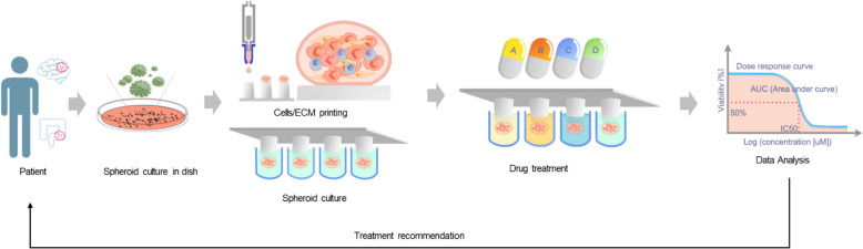
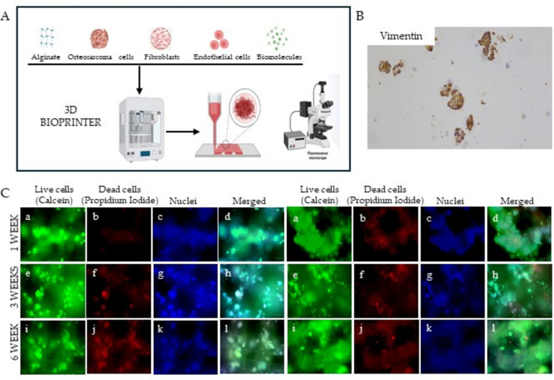
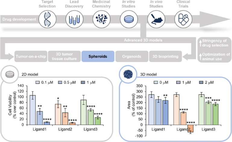
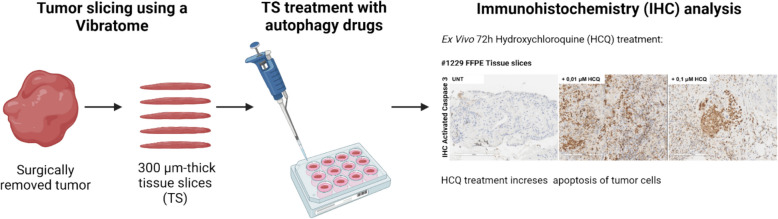
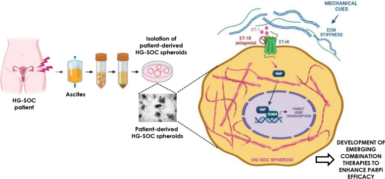
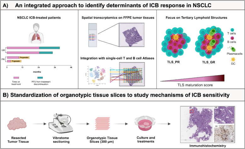
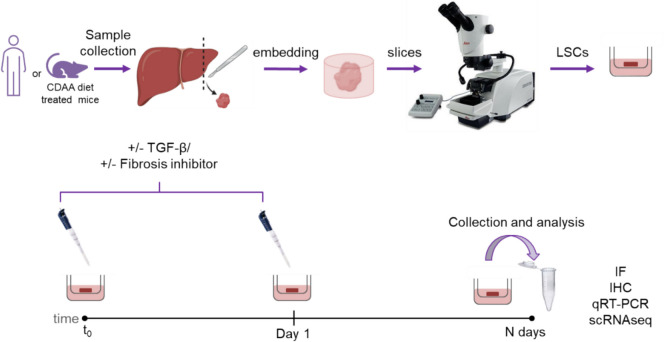
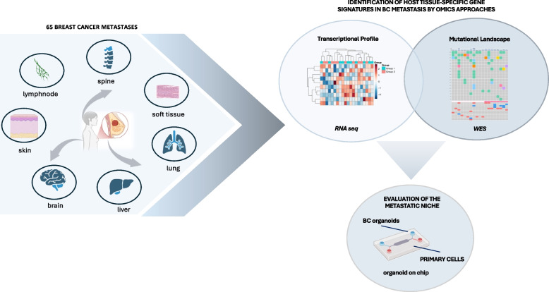
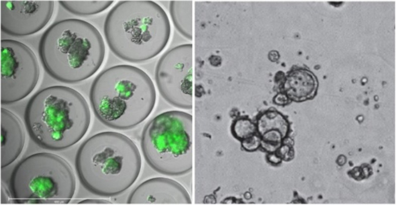
On September 23-24 (2024) the 6th Workshop IRE on Translational Oncology, titled "Cancer Organoids as Reliable Disease Models to Drive Clinical Development of Novel Therapies," took place at the IRCCS Regina Elena Cancer Institute in Rome. This prominent international conference focused on tumor organoids, bringing together leading experts from around the world.A central challenge in precision oncology is modeling the dynamic tumor ecosystem, which encompasses numerous elements that evolve spatially and temporally. Patient-derived 3D culture models, including organoids, explants, and engineered or bioprinted systems, have recently emerged as sophisticated tools capable of capturing the complexity and diversity of cancer cells interacting within their microenvironments. These models address critical unmet needs in precision medicine, particularly in aiding clinical decision-making. The rapid development of these human tissue avatars has enabled advanced modeling of cellular alterations in disease states and the screening of compounds to uncover novel therapeutic pathways.Throughout the event, distinguished speakers shared their expertise and research findings, illustrating how organoids are transforming our understanding of treatment resistance, metastatic dynamics, and the interaction between tumors and the surrounding microenvironment.This conference served as a pivotal opportunity to strengthen international collaborations and spark innovative translational approaches. Its goal was to accelerate the shift from preclinical research to clinical application, paving the way for increasingly personalized and effective cancer therapies.

Keywords: Cancer spheroid; Organoid; Patient-derived 3D culture model; Precision medicine; Preclinical models; Targeted therapy.


Conflict of interest statement

Declarations. Ethics approval and consent to participate: Not applicable. Consent for publication: All the authors agree to publish this paper. Competing interests: The authors declare no competing interests.

Figures

Fig. 1
Fig. 1
Endometrial Cancer Organoids Workflow
Fig. 2
Fig. 2
Functional Precision Radio-oncology in HNSCC. The schematic illustrates the potential workflow of functional precision radio-oncology. Tissue samples (tumour and adjacent normal tissue) are collected during diagnostic biopsy or curative-intent surgery. Following the establishment and expansion of organoid cultures, screening is conducted for single and combination treatment regimens. Experimental treatment efficacy is assessed using read-outs such as cell viability, clonogenic survival, and/or residual DNA double-strand breaks. Drugs are ranked based on their efficacy scores in tumour and normal organoids. Top candidate therapies are subsequently reported to molecular tumour boards to inform treatment recommendations. Figure created with biorender.com
Fig. 3
Fig. 3
Workflow from high-throughput screenings in a microfluidic PDO platfoform towards single cell multi-omic analyses
Fig. 4
Fig. 4
Left panel: Schematic of patient-derived pancreatic ductal adenocarcinoma (PDAC) organoid grown within an engineered matrix (termed a HELP hydrogel). Changing the number of chemical crosslinks between the biopolymers within the HELP hydrogel creates matrices with low, medium, and high stiffness. Right panel: PDAC organoids proliferated and grew similarly well within a commercially available Cultrex matrix or the engineered HELP hydrogels of Low, Medium, and High stiffness. Organoids grown in stiffer matrices (HELP High) became less sensitive to the chemotherapy gemcitabine, as shown by less immunostaining for the apoptosis marker cleaved caspase 3. Reprinted with permission (The figure is a modified version of a Fig. 1 in reference 16)
Fig. 5
Fig. 5
The Personalized Functional Profiling (PFP) technology. Surgical patient biopsies are dissociated and culturing tumor spheroid. Dissociated cells are printed onto a 3D-pillar support system using robot technology and grown as tumor spheroids. These tumor spheroids have the same genomic composition as the tumor of origin. Tumor spheroids are then challenged by a large panel of drugs (including FDA approved drugs). Growth (size of spheroids) is measured by an automated confocal microscope. To foster a comparative analysis of drug responses, we will employ an analysis of drug sensitivity, including the half-maximal inhibitory concentration (IC50) and the area under curve (AUC) of the dose–response curve (DRC). Drugs that show efficacy can potentially be reported to patients
Fig. 6
Fig. 6
TIICs expansion and characterization. A Images representative of one Urachal carcinoma sample mPDO in brightfield and multiplex immunofluorescence (3D passage 1); depicted are cytokeration-7 (red), αSMA (green) and CD45 (magenta). B Images representative of one renal medullary carcinoma sample TIICs in brightfield and multiplex immunofluorescence (passage 1 and 3, respectively); depicted are cytokeration-7 (red), αSMA (green) CD45 (magenta). C Graphical representation of the proportion of cells positive for CD45, Vimentin and Cytokeratin within the parental tumor sample processed analysed via multiplex immunofluorescence. Coral colouring marks the tumours showing TIICs expansion. D Co-culture conditions tested for the medullary renal carcinoma cPDO with autologous PBMCs and TIICs with graphical representation of the organoid growth
Fig. 7
Fig. 7
Bladder Cancer PDOs Workflow
Fig. 8
Fig. 8
Schematic representation of FGFR2 fusion-positive intrahepatic cholangiocarcinoma (iCCA) (A) and generation of iCCA models based on murine liver organoids genetically modified to express FGFR2 fusion proteins (B)
Fig. 9
Fig. 9
A Diagram of 3D extrusion based bioprinter working process: biomaterial, populations of interest and biomolecules are inserted into the instrument and extruded together in a software-aided shape. B Immunohistochemistry analysis conducted on 3D OS bioprinted models showing the positivity for Vimentin (magnification 100X). C 3D OS models were bioprinted starting from Sa-Os cell line alone (on the left) or in combination with fibroblasts (on the right). The models were cultured in a 6-well chamber slide and stained with the live-dead assay kit (Merck). The rows show live cell OS models stained with (a, e, i) Calcein-AM, (b, f, j) propidium iodide, (c,g,k) Hoechst 33,342 and (d,h,l) merged image
Fig. 10
Fig. 10
Upper panel. Schematic representation of drug development process. Bottom panels. Left panel: 2D cell viability assays performed after 48 h of treatment with the indicated compounds, at the final concentrations of 0.1, 0.5 and 1 μM. The results are expressed as the percentage of cell viability over the untreated cells. The histogram represents the mean values ± S.D. of three independent experiments. Right panel: Time course analysis of 3D tumor spheroids growth. Two days after the plating, the spheres were treated with the indicated compound at the final concentrations of 1 and 2 μM and monitored by Incucyte® S3 Live-Cell Analysis System (Essen BioScience, Ann Arbor, MI), 4X magnification. The results are expressed as the percentage of the spheroids area upon treatment relative to their own area before the compound administration. The graphs represent the mean values ± S.E.M. of at least 4 spheres. For each graph: *p < 0.05, **p < 0.01, ***p < 0.001, ****p < 0.0001
Fig. 11
Fig. 11
Organotypic tissue slices as preclinical model to test autophagy drugs. Illustration of the experimental workflow used in the study. Immunohistochemistry of activated Caspase 3 was used as a marker of apoptotic cell death. Figure created with biorender.com
Fig. 12
Fig. 12
Graphic model depicting how in 3D high-grade serous ovarian cancer (HG-SOC) patient-derived spheroid models the ET-1/ET-1R/YAP circuit, intersecting mechanical forces generated by a stiff extracellular matrix (ECM), potentiates the HG-SOC invasive behaviour and sustains PARP inhibitors (PARPi) tolerance. Figure created with biorender.com. Abbreviations: EC_PDO: Patient-derived endometrial cancer organoids, 3D: Three-dimensional. PBMC: Peripheral blood mononuclear cells. TAMs: Tumor-associated macrophages. PDOs: Patient-derived organoids. HNSCC: Head and neck squamous cell carcinoma. ECM: Extracellular matrix
Fig. 13
Fig. 13
A Workflow of the integrated approach employed to identify determinants of ICB response. Archival tumor tissues, collected at the time of surgery, of a cohort of naïve of treatment NSCLC patients who recurred and were treated with ICB in accordance with guidelines were scored as poor (PR) and good (GR) ICB responders. Spatial transcriptomic by 10XVisium highlighted distinct composition and functionality of TLS of PR versus GR. B Standardization of organotypic tissue slices (TS) obtained from resected naïve of treatment NSCLC patients. TS were sliced, cultured, treated with ICB ex-vivo and then subjected to histological analysis. Figure created with biorender.com
Fig. 14
Fig. 14
Specimens from healthy human livers or diet-treated mice are collected and cut to obtain 250 µm thick slices (Liver Slice Cultures, LSCs) before culturing them. TGF-β is added to human LSCs while pharmacological compounds are added to mouse LSCs. Slices are collected at different time points and the expression of pro-fibrotic markers analyzed
Fig. 15
Fig. 15
Breast Cancer Metastases Workflow
Fig. 16
Fig. 16
Left, GFP-labeled MOs in micro-wells. Right, Patient-derived MOs in a well-plate, ready for drug testing

Similar articles

Cited by

References

    1. Bose S, Barroso M, Chheda MG, Clevers H, Elez E, Kaochar S, Kopetz SE, Li XN, Meric-Bernstam F, Meyer CA, et al. A path to translation: How 3D patient tumor avatars enable next generation precision oncology. Cancer Cell. 2022;40:1448–53. - PMC - PubMed
    1. de Visser KE, Joyce JA. The evolving tumor microenvironment: From cancer initiation to metastatic outgrowth. Cancer Cell. 2023;41:374–403. - PubMed
    1. Neufeld L, Yeini E, Pozzi S, Satchi-Fainaro R. 3D bioprinted cancer models: from basic biology to drug development. Nat Rev Cancer. 2022;22:679–92. - PubMed
    1. Neufeld L, Yeini E, Reisman N, Shtilerman Y, Ben-Shushan D, Pozzi S, Madi A, Tiram G, Eldar-Boock A, Ferber S, et al: Microengineered perfusable 3D-bioprinted glioblastoma model for in vivo mimicry of tumor microenvironment. Sci Adv. 2021;7(34):eabi9119. - PMC - PubMed
    1. Yeini E, Ofek P, Pozzi S, Albeck N, Ben-Shushan D, Tiram G, Golan S, Kleiner R, Sheinin R, Israeli Dangoor S, et al. P-selectin axis plays a key role in microglia immunophenotype and glioblastoma progression. Nat Commun. 1912;2021:12. - PMC - PubMed