Review
. 2025 Jan;329(1):e13431.
doi: 10.1111/imr.13431. Epub 2024 Dec 27.

Structural Immunology of SARS-CoV-2

Affiliations
Review

Structural Immunology of SARS-CoV-2

Meng Yuan et al. Immunol Rev. 2025 Jan.

Abstract

The SARS-CoV-2 spike (S) protein has undergone significant evolution, enhancing both receptor binding and immune evasion. In this review, we summarize ongoing efforts to develop antibodies targeting various epitopes of the S protein, focusing on their neutralization potency, breadth, and escape mechanisms. Antibodies targeting the receptor-binding site (RBS) typically exhibit high neutralizing potency but are frequently evaded by mutations in SARS-CoV-2 variants. In contrast, antibodies targeting conserved regions, such as the S2 stem helix and fusion peptide, exhibit broader reactivity but generally lower neutralization potency. However, several broadly neutralizing antibodies have demonstrated exceptional efficacy against emerging variants, including the latest omicron subvariants, underscoring the potential of targeting vulnerable sites such as RBS-A and RBS-D/CR3022. We also highlight public classes of antibodies targeting different sites on the S protein. The vulnerable sites targeted by public antibodies present opportunities for germline-targeting vaccine strategies. Overall, developing escape-resistant, potent antibodies and broadly effective vaccines remains crucial for combating future variants. This review emphasizes the importance of identifying key epitopes and utilizing antibody affinity maturation to inform future therapeutic and vaccine design.

Keywords: COVID‐19; SARS‐CoV‐2; neutralizing antibody; spike; vaccine; viral evolution.


Conflict of interest statement

The authors declare no conflicts of interest.

Figures

FIGURE 1
FIGURE 1
Overall structure of the SARS‐CoV‐2 spike (S) protein. (A) Schematic representation of the SARS‐CoV‐2 S protein. (B) A prefusion state of the SARS‐CoV‐2 S protein with one RBD in the “up” state (PDB 7N1Q [2]). The other two protomers with RBDs in “down” state are represented by a more faded surface. The color scheme of the highlighted regions is the same as in panel A. (C) Phylogenetic tree of coronaviruses. The phylogenetic tree was calculated by Clustal Omega [3] and edited by Interactive Tree Of Life (iTOL) [4].
FIGURE 2
FIGURE 2
Evolution of the SARS‐CoV‐2 S protein. (A) Frequencies of SARS‐CoV‐2 variants since the beginning of the COVID‐19 pandemic (colored by Pangolin Pango Lineage [13]) as calculated by Nextstrain [14] using data from GISAID [15]. (B) Prevalent mutations of SARS‐CoV‐2 variants as calculated by Outbreak.info [16]. Mutations with > 75% prevalence are shown here. RBS residues (defined as buried surface area [BSA] > 0 Å2 as calculated by PISA [Proteins, Interfaces, Structures and Assemblies]) [17] are highlighted with asterisks. (C) Increasing number of mutations on the SARS‐CoV‐2 S protein during virus evolution. Mutated residues are represented by red spheres.
FIGURE 3
FIGURE 3
Antibody sites and conservation of the SARS‐CoV‐2 S protein. Sequence conservation of SARS‐CoV‐2 variants are mapped on a prefusion S spike and its domains (PDB 7N1Q [2]) by ConSurf [23]. The color coding is consistent throughout this figure. (A) Sequence conservation of SARS‐CoV‐2 variants plotted on a SARS‐CoV‐2 S structure. B‐cell epitopes on S2 stem and fusion peptide are usually cryptic in the prefusion state and the epitope residues highlighted as red sticks in the boxes on the side. (B–E) Antibodies targeting different epitopes on the receptor‐binding domain (RBD) are classified as RBS‐A, ‐B, ‐C, ‐D, CR3022 cryptic site, N343 glycan site, and site V, which are indicated in the panels that present different views of the RBD. (F) Antibodies targeting different epitopes on the N‐terminal domain (NTD).
FIGURE 4
FIGURE 4
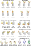
Public classes of antibodies targeting the SARS‐CoV‐2 S protein. Antibody/antigen complex structures are from the Protein Data Bank (PDB) with the heavy and light chains of the antibodies shown in orange and yellow, respectively. The antigens are shown in transparent gray for RBD and NTD, and as black tubes for the S2 stem helices. The images of the negative stain EM maps of the anti‐S2‐apex antibodies are from the Electron Microscopy Data Bank (EMDB). High‐resolution structures of these antibodies are not available.
FIGURE 5
FIGURE 5
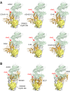
Antibodies targeting the conserved CR3022 cryptic site that clash with ACE2 binding. (A) Structurally convergent public antibodies with the “YYDxxG” motif in the CDR H3 target the conserved CR3022 cryptic site, while their angles of approach enable blocking of binding to angiotensin‐converting enzyme 2 (ACE2). (B) Antibodies targeting the RBS‐D/CR3022 (Class 1/4) site. The epitopes span from a corner of the RBS to the conserved CR3022 cryptic site. The SARS‐CoV‐2 RBD is shown as a white surface with the CR3022 site highlighted in yellow, while the variable domains of the antibodies are in orange. The RBD–antibody structure is superimposed onto an RBD‐ACE2 structure (PDB 6M0J) and illustrates that the antibodies would clash (red arrows) with ACE2 (green). The PDB codes of the antibody/antigen complex structures shown here are: COVA1‐16: 7JMW; ADI‐62113: 7T7B; 10–40: 7SD5; C022: 7RKU; 2–36: 7N5H; CC25.54: 8SIR; ADG20: 7U2D; SC27: 8VIF; SA55: 7Y0W.
FIGURE 6
FIGURE 6
Sequence alignment of the fusion peptide, S2 apex, and S2 stem regions across coronaviruses. Amino acids in blue boxes are positions which have a single, fully conserved residue, while cyan boxes indicate conservation between groups of similar properties (scoring > 0 in the Gonnet PAM 250 matrix) [183]. The sequence alignment was performed using Clustal Omega [3]. The fusion peptide and S2 apex are conserved across all genera of coronaviruses, while the S2 stem region is conserved across betacoronaviruses where sequence conservation colors are only shown for the betacoronaviruses.
FIGURE 7
FIGURE 7
An overview of the breadth and potency of mAbs targeting the SARS‐CoV‐2 S protein. The neutralizing IC50 values are to the wild‐type SARS‐CoV‐2 from the original papers of each mAbs and shown along the y‐axis. The reactive breadth of binding on the x‐axis includes cross‐reactivity to SARS‐CoV‐2 variants, sarbecoviruses, betacoronaviruses, and other genera of coronaviruses. Note that neutralizing IC50 values are from different groups and are only comparable in terms of order of magnitude.
FIGURE 8
FIGURE 8
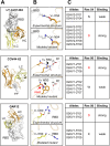
Allelic polymorphisms impact the binding activity of antibodies to SARS‐CoV‐2 antigens. (A) Overall view of antibody/antigen interactions. (B) Detailed interactions between the allelic polymorphic residues and the antigen, as well as the modeled allelic mutations. (C) Allele usage of the paratopic residues. Residues that correspond to the representative antibody in panel A are shown in red. The information of alleles is from the IMGT Repertoire [220]. Binding affinities of the wild‐type and mutated antibodies to SARS‐CoV‐2 S were measured experimentally and reported in our previous studies [61, 218]. Three representative anti‐SARS‐CoV‐2 antibodies with allelic polymorphisms LY‐CoV1404, COV44‐62, and GAR12 are shown here. Other cases are comprehensively analyzed and reported in our previous study [218]. Antibody heavy and light chains are shown in orange and yellow, respectively. Antigens are shown in white. Allelic polymorphic residues are labeled. Red disks indicate significant van der Waals overlap (distance < 2.8 Å), hence representing a steric clash. H bonds and salt bridges are represented by black dashed lines. Kabat numbering is applied to all antibodies. Allelic mutations are modeled by FoldX [221].

Similar articles

  • Escape from neutralizing antibodies by SARS-CoV-2 spike protein variants.
    Weisblum Y, Schmidt F, Zhang F, DaSilva J, Poston D, Lorenzi JC, Muecksch F, Rutkowska M, Hoffmann HH, Michailidis E, Gaebler C, Agudelo M, Cho A, Wang Z, Gazumyan A, Cipolla M, Luchsinger L, Hillyer CD, Caskey M, Robbiani DF, Rice CM, Nussenzweig MC, Hatziioannou T, Bieniasz PD. Weisblum Y, et al. Elife. 2020 Oct 28;9:e61312. doi: 10.7554/eLife.61312. Elife. 2020. PMID: 33112236 Free PMC article.
  • Pan-beta-coronavirus subunit vaccine prevents SARS-CoV-2 Omicron, SARS-CoV, and MERS-CoV challenge.
    Wang G, Verma AK, Guan X, Bu F, Odle AE, Li F, Liu B, Perlman S, Du L. Wang G, et al. J Virol. 2024 Sep 17;98(9):e0037624. doi: 10.1128/jvi.00376-24. Epub 2024 Aug 27. J Virol. 2024. PMID: 39189731 Free PMC article.
  • Structural and antigenic variations in the spike protein of emerging SARS-CoV-2 variants.
    Mittal A, Khattri A, Verma V. Mittal A, et al. PLoS Pathog. 2022 Feb 17;18(2):e1010260. doi: 10.1371/journal.ppat.1010260. eCollection 2022 Feb. PLoS Pathog. 2022. PMID: 35176090 Free PMC article. Review.
  • SARS-CoV-2 RBD antibodies that maximize breadth and resistance to escape.
    Starr TN, Czudnochowski N, Liu Z, Zatta F, Park YJ, Addetia A, Pinto D, Beltramello M, Hernandez P, Greaney AJ, Marzi R, Glass WG, Zhang I, Dingens AS, Bowen JE, Tortorici MA, Walls AC, Wojcechowskyj JA, De Marco A, Rosen LE, Zhou J, Montiel-Ruiz M, Kaiser H, Dillen JR, Tucker H, Bassi J, Silacci-Fregni C, Housley MP, di Iulio J, Lombardo G, Agostini M, Sprugasci N, Culap K, Jaconi S, Meury M, Dellota E Jr, Abdelnabi R, Foo SC, Cameroni E, Stumpf S, Croll TI, Nix JC, Havenar-Daughton C, Piccoli L, Benigni F, Neyts J, Telenti A, Lempp FA, Pizzuto MS, Chodera JD, Hebner CM, Virgin HW, Whelan SPJ, Veesler D, Corti D, Bloom JD, Snell G. Starr TN, et al. Nature. 2021 Sep;597(7874):97-102. doi: 10.1038/s41586-021-03807-6. Epub 2021 Jul 14. Nature. 2021. PMID: 34261126 Free PMC article.
  • Comprehensive Overview of Broadly Neutralizing Antibodies against SARS-CoV-2 Variants.
    Cui L, Li T, Xue W, Zhang S, Wang H, Liu H, Gu Y, Xia N, Li S. Cui L, et al. Viruses. 2024 Jun 1;16(6):900. doi: 10.3390/v16060900. Viruses. 2024. PMID: 38932192 Free PMC article. Review.

Cited by

References

    1. WHO , “WHO COVID‐19 Dashboard,” https://data.who.int/dashboards/covid19/cases.
    1. Cai Y., Zhang J., Xiao T., et al., “Structural Basis for Enhanced Infectivity and Immune Evasion of SARS‐CoV‐2 Variants,” Science 373, no. 6555 (2021): 642–648. - PMC - PubMed
    1. Sievers F., Wilm A., Dineen D., et al., “Fast, Scalable Generation of High‐Quality Protein Multiple Sequence Alignments Using Clustal Omega,” Molecular Systems Biology 7 (2011): 539. - PMC - PubMed
    1. Letunic I. and Bork P., “Interactive Tree of Life (iTOL): An Online Tool for Phylogenetic Tree Display and Annotation,” Bioinformatics 23, no. 1 (2007): 127–128. - PubMed
    1. Shi X., Li Y., Yan L., Yang G., and Qiang M., “Neutralizing Antibodies Targeting SARS‐CoV‐2 Spike Protein,” Stem Cell Research 50 (2020): 102125. - PMC - PubMed

Publication types

MeSH terms