

Comparison of Marker-Based RSA and CT-RSA for Analyzing Micromotions After Distal Radius Osteotomy: A 1-Year Retrospective Study of 24 Patients
- PMID: 39731268
- PMCID: PMC11806653
- DOI: 10.1002/jor.26031
Comparison of Marker-Based RSA and CT-RSA for Analyzing Micromotions After Distal Radius Osteotomy: A 1-Year Retrospective Study of 24 Patients
Abstract
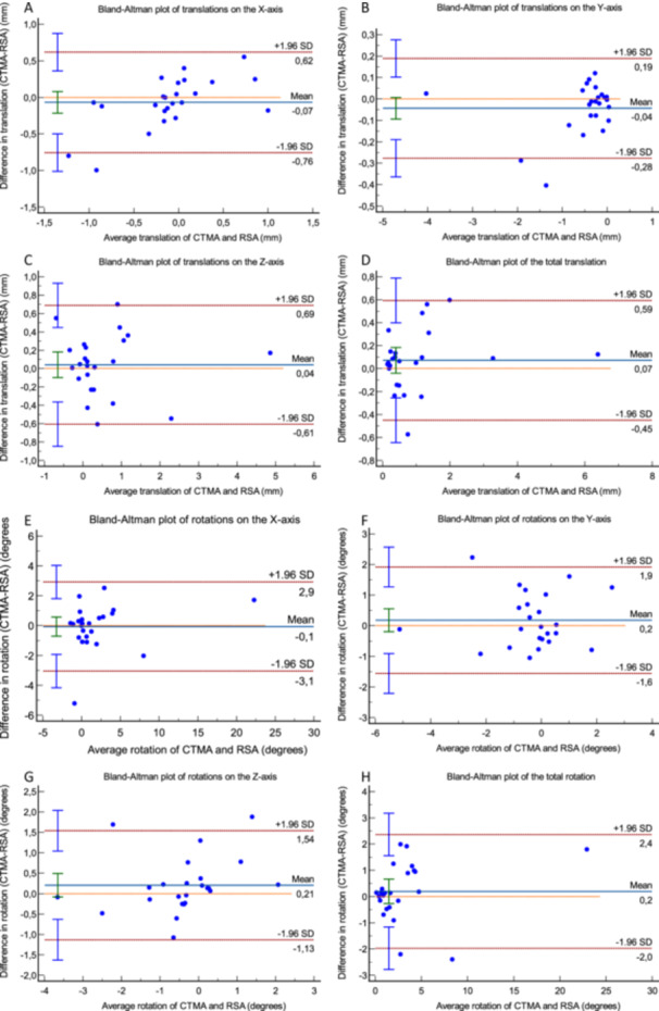
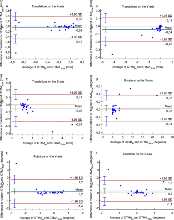
Radiostereometric Analysis (RSA) is the most accurate method for determining early micromotions of orthopedic implants. Computed Tomography Radiostereometric Analysis (CT-RSA) is a method that can be used to determine implant and bone micromovements using low-dose CT scans. This study aimed to evaluate the reliability of the CT-RSA method in measuring the interfragmental mobility in patients who have undergone a correction osteotomy due to a malunited distal radius fracture. Twenty-four patients were included and operated with a radiolucent volar plate. Markers were embedded in the plate and bone. RSA and CT examinations were obtained postoperatively up to 1-year postoperative. Micromovements of the distal radius segment relative to the proximal were compared between the methods with paired analysis and Bland-Altman plots. The limits of clinical significance were: dorsal/volar tilt < 10°, radial shortening < 5 mm, radial inclination ≥ 15°, and radial shift < 5 mm. For the dorsal/volar tilt, the paired analysis between the two methods, showed a mean difference (95% CI) of -0.06° (-0.67 to 0.55), for radial compression-0.04 mm (-0.09 to 0.01), for radial inclination 0.21° (-0.06 to 0.48), and for radial shift -0.07 mm (-0.21 to 0.07). The paired analysis for micromotions showed that the thresholds of clinical significance are excluded from the difference's 95% CI. The Bland-Altman plots showed comparable results up to 1 year, considering clinically relevant thresholds. In conclusion, the CT-RSA method is comparable to that of marker-based RSA in measuring micromotions after wrist osteotomy, as the differences between the methods are not clinically significant.
Keywords: CT‐based; computed tomography; distal radius; micromotions; radiostereometric analysis.
© 2024 The Author(s). Journal of Orthopaedic Research® published by Wiley Periodicals LLC on behalf of Orthopaedic Research Society.
Conflict of interest statement
Olof Sandberg works as an engineer and researcher at Sectra, the company that owns CTMA, the CT‐RSA analysis software that was used in this study, which could imply conflict of interest, but he had no part in the clinical interpretation of the results. The other authors declare no conflicts of interest.
Figures
References
-
- Leong N. L., Buijze G. A., Fu E. C., Stockmans F., Jupiter J. B., and Distal Radius Malunion (DiRaM) Collaborative Group , “Computer‐Assisted Versus Non‐Computer‐Assisted Preoperative Planning of Corrective Osteotomy for Extra‐Articular Distal Radius Malunions: A Randomized Controlled Trial,” BMC Musculoskeletal Disorders 11, no. 1 (2010): 282, 10.1186/1471-2474-11-282/FIGURES/2. - DOI - PMC - PubMed
Publication types
MeSH terms
LinkOut - more resources
Full Text Sources
Medical
