

Impact of adipocytes on ultrasound evaluation of parathyroid adenomas
- PMID: 39731664
- PMCID: PMC12018633
- DOI: 10.1007/s10396-024-01511-2
Impact of adipocytes on ultrasound evaluation of parathyroid adenomas
Abstract
Purpose: Parathyroid lipoadenomas are difficult to recognize preoperatively; hence, they may remain undetected. Difficulty in recognition is thought to be due to the adipocytes present in the tumor. This study aimed to clarify the impact of adipocytes as a component of parathyroid adenomas on ultrasound evaluation.
Methods: Eighteen parathyroid adenoma cases, in which the adipose tissue accounted for more than 10% of the tumors, were included in this study. Of these, five were consistent with lipoadenomas. Twenty-five consecutive patients with parathyroid adenoma without adipocytes were used as controls.
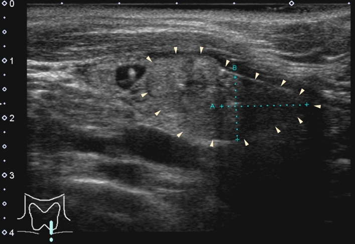
Results: Ultrasonography revealed a lipoadenoma detection rate of 20.0%. This increased to 80.0% at re-examinations performed after obtaining information from other imaging modalities. Compared with parathyroid adenoma cases with no adipocytes or few adipocytes, the frequencies of ill-defined margins, iso- and/or hyperechogenicity, heterogeneous consistency with a two-tone pattern, poor vascular flow, no polar artery, and no hyperechoic line were significantly higher in parathyroid lipoadenoma cases. The hyperechoic and isoechoic areas in tumors with a two-tone pattern correspond to adipocyte- and parathyroid cell-rich areas, respectively. The lipoadenoma tumor sizes measured using ultrasound tended to be smaller than the actual sizes.
Conclusions: The characteristic ultrasound findings of lipoadenomas were clearly different from those of parathyroid adenomas with or without adipocytes. We believe that our findings may contribute to an increased detection rate of lipoadenomas and allow us to consider them in the differential diagnosis.
Keywords: Lipoadenoma; Parathyroid adenoma; Two-tone pattern; Ultrasound.
© 2024. The Author(s).
Conflict of interest statement
Declarations. Conflict of interest: The authors declare no conflicts of interest. Ethical approval: The study protocol was reviewed and approved by the Institutional Review Board of Kuma Hospital (approval number: 20231012–4), and was conducted in accordance with the 1964 Declaration of Helsinki and its amendments or comparable ethical standards.
Figures


Similar articles
-
A rare cause of primary hyperparathyroidism: Parathyroid lipoadenoma.Auris Nasus Larynx. 2018 Dec;45(6):1245-1248. doi: 10.1016/j.anl.2018.05.001. Epub 2018 May 16. Auris Nasus Larynx. 2018. PMID: 29778311
-
Parathyroid lipoadenomas: a rare cause of primary hyperparathyroidism.Endocr Pract. 2006 Mar-Apr;12(2):131-6. doi: 10.4158/EP.12.2.131. Endocr Pract. 2006. PMID: 16690459
-
Pathology quiz case 2. Parathyroid lipoadenoma.Arch Otolaryngol Head Neck Surg. 2006 Dec;132(12):1391-3. doi: 10.1001/archotol.132.12.1391. Arch Otolaryngol Head Neck Surg. 2006. PMID: 17178956 No abstract available.
-
Functioning lipoadenoma of the parathyroid: case report and literature review.Mt Sinai J Med. 1989 Mar;56(2):114-7. Mt Sinai J Med. 1989. PMID: 2664480 Review.
-
The elusive parathyroid adenoma: techniques for detection.Ultrasound Q. 2013 Sep;29(3):179-87. doi: 10.1097/RUQ.0b013e3182a1ba6f. Ultrasound Q. 2013. PMID: 23975046 Review.
References
-
- Hyrcza MD, Sargın P, Mete O. Parathyroid lipoadenoma: A clinicopathological diagnosis and possible trap for the unaware pathologist. Endocr Pathol. 2016;27:34–41. - PubMed
-
- Onoda N, Hirokawa M, Miya A, et al. Lipoadenoma of the parathyroid: Characteristics of a rare cause of hyperparathyroidism. Endocr J. 2022;69:1227–32. - PubMed
-
- Özden S, Güreşci S, Saylam B, et al. A rare cause of primary hyperparathyroidism: Parathyroid lipoadenoma. Auris Nasus Larynx. 2018;45:1245–8. - PubMed
-
- Erickson LA, Mete O, Juhlin CC, et al. Overview of the 2022 WHO classification of parathyroid tumors. Endocr Pathol. 2022;33:64–89. - PubMed
MeSH terms
LinkOut - more resources
Full Text Sources
