. 2025 Feb 20;32(2):307-324.e15.
doi: 10.1016/j.chembiol.2024.12.001. Epub 2024 Dec 27.

Discovery of a Pseudomonas aeruginosa-specific small molecule targeting outer membrane protein OprH-LPS interaction by a multiplexed screen

Affiliations

Discovery of a Pseudomonas aeruginosa-specific small molecule targeting outer membrane protein OprH-LPS interaction by a multiplexed screen

Bradley E Poulsen et al. Cell Chem Biol. .

Abstract

The surge of antimicrobial resistance threatens efficacy of current antibiotics, particularly against Pseudomonas aeruginosa, a highly resistant gram-negative pathogen. The asymmetric outer membrane (OM) of P. aeruginosa combined with its array of efflux pumps provide a barrier to xenobiotic accumulation, thus making antibiotic discovery challenging. We adapted PROSPECT, a target-based, whole-cell screening strategy, to discover small molecule probes that kill P. aeruginosa mutants depleted for essential proteins localized at the OM. We identified BRD1401, a small molecule that has specific activity against a P. aeruginosa mutant depleted for the essential lipoprotein, OprL. Genetic and chemical biological studies identified that BRD1401 acts by targeting the OM β-barrel protein OprH to disrupt its interaction with LPS and increase membrane fluidity. Studies with BRD1401 also revealed an interaction between OprL and OprH, directly linking the OM with peptidoglycan. Thus, a whole-cell, multiplexed screen can identify species-specific chemical probes to reveal pathogen biology.

Keywords: LPS; OprH; Pseudomonas aeruginosa; Tol-Pal; chemical screening; gram-negative bacteria; growth inhibitor; lipopolysaccharide; outer membrane.


Conflict of interest statement

Declaration of interests The authors declare no competing interests.

Figures

Figure 1.
Figure 1.. Genetic depletion strains to increase small molecule sensitivity.
(A) P. aeruginosa essential OMPs and OMAPs targeted in this study. Structures of P. aeruginosa BamE (7JRK), LolA (2W7Q), LptDE (5IVA) and LptA (4UU4) from Protein Data Bank; all others are AlphaFold predictions. (B) Gene of interest (light grey) driven by one of eight constitutive promoters (PPa1–8) was cloned into the mini-Tn7 plasmid with a unique barcode (BC, green), integrated into P. aeruginosa at a single conserved attTn7 site (blue), and the native gene copy (dark grey) was replaced by kanamycin resistance gene (red). (C) Relative expression strengths of promoter set. The eight constitutive promoters (PPa1–8) used for creating depletion strains were each integrated at the attTn7 site driving GFP expression in P. aeruginosa demonstrating varying expression levels as measured by GFP fluorescence. E. coli consensus promoter (PEc) and IPTG-inducible lac promoter (Plac) ± inducer are shown for comparison. Values are subtracted from the wild-type PA14 background (n=4). (D) Growth kinetic of P. aeruginosa depletion strains (black or colored lines) and the wild-type strain PA14 (red) measured by an absorbance-based assay (OD600nm; mean shown, n=16). All strains except for bamE- and lptD-hypomorphs grew with similar rates. oprL- and tolB-hypomorph strains grew to a lower final OD600nm. (E) Impact of MurA expression on fosfomycin activity measured by OD600nm -based growth assay. Five murA-hypomorph strains (with PPa3–7 promoters) exposed to a dose response of fosfomycin, a MurA inhibitor, for 16 hours (n=3). (F) Impact of LptD downregulation on POL7080 activity. OD600nm of lptD-hypomorph strains (with PPa6 or PPa7 promoters) after exposure to a dose response of POL7080 for 16 hours (n=3). Wild-type PA14 strain is highlighted in red in (E) and (F). Error bars indicate SEM. See also Figure S1A and Data S1-2 and S6-7.
Figure 2.
Figure 2.. Development of a multiplexed chemical screening assay.
(A) Multiplexed screening strategy. 76 bp DNA barcodes are chromosomally integrated into each species and hypomorph strain. Strains are individually grown to mid-log phase, pooled, and dispensed to 384-well plates ± compound. After incubation with a chemical library, cells are lysed, and lysate is added to PCR reactions in 384-well plates to amplify barcoded region. PCR products are pooled, ligated with Illumina adaptors, sequenced via Illumina, and plate-well indices are demultiplexed to determine the read count of each strain/well. (B) Mean read counts of each barcoded species is proportional to OD600nm in an evenly mixed pool of the 5 species (n=12; error bars indicate SD). (C) Growth, as reflected in log2 fold-change of sequenced barcode reads (relative to DMSO), of wild-type bacterial (colored circles) and P. aeruginosa depletion strains (grey circles) grown in multiplex for 12 hours, in response to known antibacterial compounds (n=12). The broad-spectrum antibiotic ciprofloxacin (CIP, 8 μM) inhibits growth for all strains; trimethroprim (TMP, 32 μM) and POL7080 (High, 64 nM; Low, 8 nM) inhibit only select strains and species. The target of TMP, FolA, and the target of POL7080, LptD, are indicated. At low [POL7080], other hypomorphs related to LPS synthesis/transport, e.g., lpxC and lptA, are also depleted. See also Figures S1B-D, Table S2 and Data S3-7.
Fig. 3.
Fig. 3.. SLF metric from primary multiplexed screen validated as a measure of P. aeruginosa strain sensitivity to a given compound.
(A) Standardized log fraction (SLF), a measure of the fractional composition of a given strain in a well relative to its fraction in vehicle-control (DMSO) wells. When a compound treatment specifically inhibits growth of a particular strain, its SLF value becomes negative. When a compound non-specifically inhibits growth of all strains, the SLF value remains near zero because the fraction of the population taken up by a given strain is like a DMSO well, even though the number of read counts diminishes. LFAC is the log10 of the fraction of a strain in a well, with A representing strain A, and C representing the compound. (B) For each compound-strain interaction tested in the demultiplexed assay, the median % growth inhibition measured by OD600nm, is plotted against its corresponding SLF from the primary multiplexed screen. SLF values are grouped into bins, with each bin representing all SLF values its label and the next lower value (e.g., bin “–5” indicates –6 ≤ SLF < –5). N=number of compound-strain pairs included in each bin. Increasingly negative SLF values in the multiplexed screen correspond to increasing growth inhibition when the strain is individually tested against the compound in the OD600nm assay. (C) BRD1401, a compound that was specifically active against P. aeruginosa oprL-hypomorph strain in the multiplexed screen and whose activity was confirmed in the demultiplexed assay. Median % growth inhibition of BRD1401 in the demultiplexed assay versus its SLF in the multiplexed screen is plotted. Each point represents a different P. aeruginosa strain. See also Figure S1E and Tables S3-S4.
Figure 4.
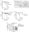
Figure 4.. BRD1401 has specific activity towards P. aeruginosa when OprL or TolB is depleted.
(A) Correctly assigned structure of BRD1401 and 5 synthesized analogs. Growth inhibitory activity against the oprL-KD strain, measured as MIC50 in an absorbance (OD600nm)-based growth, is shown. Activity of BRD1401 towards (B) bacterial species other than P. aeruginosa that were included in the multiplexed screen, P. aeruginosa PA14 and the oprL-hypomorph strains, (C) P. aeruginosa wild-type PA14 and a select subset of hypomorph strains, and (D) the oprL-KD strain in a demultiplexed OD600nm-based growth assay. Normalized growth is plotted against BRD1401 concentration (n=3, error bars indicate SD). BRD1401 does not inhibit other bacterial species while it shows specific inhibition of P. aeruginosa strains depleted of OprL or TolB. In the oprL-KD strain, an arabinose dose-dependent activity of BRD1401 is observed. oprL expression is under the control of the arabinose-inducible ParaBAD promoter in this strain. See also Figures S2-S3, S6H and Data S1-2 and S6-7.
Figure 5.
Figure 5.. OprH is necessary for BRD1401 activity.
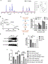
BRD1401 activity towards (A) the ΔoprL-MW strain and the isogenic wild-type strain (WT), (C) the parent oprL-hypomorph strain and the three resistant clones, R9, R10 and R11, and (D) the oprL-KD and oprL-KDΔoprH strains, as measured by an absorbance (OD600nm)-based growth assay. Normalized growth is plotted against BRD1401 concentration (n=3, error bars indicate SD). BRD1401 was active (A) in the absence of OprL, but (D) required OprH. oprL-KD and oprL-KDΔoprH strains were grown in medium with 0.063% arabinose, which regulates OprL expression through the arabinose-inducible ParaBAD promoter. (B) Table listing mutations identified in the oprH-phoPQ operon by whole-genome sequencing of the three BRD1401-resistat oprL-hypomorph clones. SNP indicates the single nucleotide polymorphism detected and AA indicates the mutated amino acid residue. (C) All three clones showed high level resistance. (E) BRD1401 is active towards intracellular P. aeruginosa. Shown is % survival, calculated from colony forming units and normalized to DMSO vehicle control, of P. aeruginosa PAO1 strain recovered from bladder epithelial cells after treatment for 24 or 48 hours with indicated concentrations of BRD1401 (n=3, error bars indicate SEM). *, p <.05; **, p <.005; unpaired Student’s parametric t-test with Welch’s correction. See also Figure S5 and Data S7.
Figure 6.
Figure 6.. BRD1401 activity is linked to OprH binding and functional inhibition.
(A) BRD1401 binds to OprH. Overlaid 1H-spectra of the aromatic region chemical shifts and intensities of 500 μM 1401-C solution (left) in the presence of 15 μM P. aeruginosa OprH (right; Method Details) without irradiation of OprH (black) and with irradiation of OprH (blue), inducing a decrease in 1401-C signal intensity due to a negative Ligand-Protein distance-dependent Nuclear Overhauser Effect (NOE). The difference spectrum (STD in red) between black and blue spectra corresponds to the magnitude of the NOE indicating binding dependent loss of 1401-C signal in presence of OprH. Calculated %STD for 1401-C analog with and without OprH is plotted in the box. (B) OprH binding correlates with BRD1401 activity. Growth inhibitory activity towards oprL-KD strain, measured as IC50, of BRD1401 and 5 analogs is inversely correlated with the ΔSTD binding parameters (Method Details) from STD-NMR with OprH protein. Fit is simple linear regression; data represents 2 independent experiments for each analog. (C) BRD1401 decreases LPS binding to OprH in vitro measured by a trypsin digestion protection assay (see schematic and Method Details). Band intensity, normalized to vehicle control (dashed line = 1), of the LPS-protected ~14.5 kDa band is plotted after exposure of OprH protein to a panel of compounds [BRD1401 and 1401-C, which are active; the inactive analog IA-1; and positive control polymyxin B (PMB)] and Kdo2-Lipid A (n=2 or 3). *, p <.05; **, p <.01; one sample t-test in comparison to the vehicle control. (D-E) BRD1401 decreases LPS binding to OprH in the OM of P. aeruginosa. StrepII-tagged OprH was pulled down from the lysates of the oprL-KDΔoprH/pRha-StrepII-oprH strain exposed to vehicle-control (DMSO), BRD1401 at 7.8 or 15.6 μM, or the inactive analog (IA-1) at 62.5 μM, followed by western blotting with anti-LPS-O10 and anti-StrepII antibodies (Method Details). Shown is the ratio of LPS band intensity relative to OprH band intensity in the elution samples, quantified by Fiji (ImageJ) and normalized to vehicle control sample (n=3). *, p <.05; unpaired Student’s parametric t-test with Welch’s correction. (F) BRD1401 increases membrane fluidity. Wild-type PA14, PA14ΔoprH, or oprL-hypomorph strain grown in media with the indicated concentrations of salt (NaCl) and exposed to 250 μM BRD1401 (1401) where indicated. Cultures were then treated with a pyrene dye and processed to measure relative fluidity normalized to PA14 strain grown with no salt (Method Details; n=6–18 biological replicates). High salt decreases membrane fluidity, while OprH deletion, OprL depletion and BRD1401 increase membrane fluidity. *, p <.05; ****, p <.0001; unpaired Student’s parametric t-test with Welch’s correction. Error bars indicate SEM. See also Figure S6 and Data S7.
Figure 7.
Figure 7.. OprH binds to OprL.
OprL binding to OprH is specific. (A) Western blot detection of OprL-FLAG and StrepII-OprH in the oprL-FLAG-KDStrepII-oprH/pRha-oprH strain after cell lysis (LYSATE) and after pulldown and elution (ELUTE) (representative blot is shown). Strain was exposed to varying doses of rhamnose in the medium to drive the expression of untagged OprH, while OprL-FLAG expression was induced with 0.5% arabinose and StrepII-OprH expression was driven by the native promoter. OprL and OprH were detected with anti-FLAG and anti-StrepII antibodies, respectively. RpoA detected by anti-RpoA antibody was the loading control. (B) Quantification of the western blot assay shown in (A) using Fiji (ImageJ; Method Details). Ratio of OprL-FLAG:StrepII-OprH band intensities (first normalized to StrepII-OprH band intensity and then further normalized to the control sample (0% rhamnose; dashed line = 1) is plotted. With increasing concentrations of rhamnose resulting in increasing expression of free, untagged OprH to compete with StrepII-OprH, the ratio of OprL-FLAG to StrepII-OprH decreased. *, p <.05; one sample t-test in comparison to the 0% rhamnose control sample. (C) Proposed model for MOA of BRD1401. OprL depletion and high salt induces PhoPQ-OprH expression and alters membrane fluidity, with the addition of BRD1401 disrupting LPS-OprH interaction by binding OprH and increasing membrane fluidity followed by cell death. OprH is necessary for BRD1401 activity with oprH deletion conferring resistance leading to cell survival. Created with BioRender.com. See also Figure S7 and Data S7.

Update of

References

    1. Johnson EO, LaVerriere E, Office E, Stanley M, Meyer E, Kawate T, Gomez JE, Audette RE, Bandyopadhyay N, Betancourt N, et al. (2019). Large-scale chemical-genetics yields new M. tuberculosis inhibitor classes. Nature 571, 72–78. 10.1038/s41586-019-1315-z. - DOI - PubMed
    1. Brown ED, and Wright GD (2016). Antibacterial drug discovery in the resistance era. Nature 529, 336–343. 10.1038/nature17042. - DOI - PubMed
    1. Silver LL (2011). Challenges of antibacterial discovery. Clin Microbiol Rev 24, 71–109. 10.1128/CMR.00030-10. - DOI - PMC - PubMed
    1. Tacconelli E, Carrara E, Savoldi A, Harbarth S, Mendelson M, Monnet DL, Pulcini C, Kahlmeter G, Kluytmans J, Carmeli Y, et al. (2018). Discovery, research, and development of new antibiotics: the WHO priority list of antibiotic-resistant bacteria and tuberculosis. Lancet Infect Dis 18, 318–327. 10.1016/S1473-3099(17)30753-3. - DOI - PubMed
    1. Payne DJ, Gwynn MN, Holmes DJ, and Pompliano DL (2007). Drugs for bad bugs: confronting the challenges of antibacterial discovery. Nat Rev Drug Discov 6, 29–40. 10.1038/nrd2201. - DOI - PubMed

MeSH terms

Substances

LinkOut - more resources