. 2024 Dec 28;24(1):427.
doi: 10.1186/s12906-024-04742-5.

The ability of clostridium novyi-NT spores to induce apoptosis via the mitochondrial pathway in mice with HPV-positive cervical cancer tumors derived from the TC-1 cell line

Affiliations

The ability of clostridium novyi-NT spores to induce apoptosis via the mitochondrial pathway in mice with HPV-positive cervical cancer tumors derived from the TC-1 cell line

Behrouz Ebadi Sharafabad et al. BMC Complement Med Ther. .

Abstract

Background: A precise observation is that the cervix's solid tumors possess hypoxic regions where the oxygen concentration drops below 1.5%. Hypoxia negatively impacts the host's immune system and significantly diminishes the effectiveness of several treatments, including radiotherapy and chemotherapy. Utilizing oncolytic spores of Clostridium novyi-NT to target the hypoxic regions of solid tumors has emerged as a noteworthy treatment strategy.

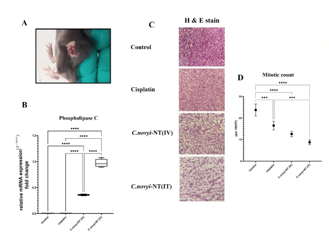
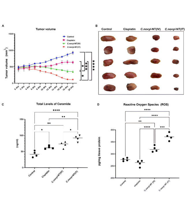
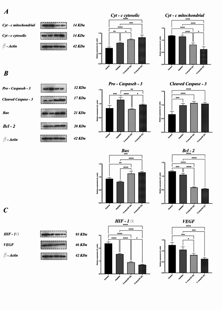
Methods: The transplantation procedure involved injecting TC-1 cells, capable of expressing HPV-16 E6/7 oncoproteins, into the subcutaneous layer of 6-8-week-old female C57/BL6 mice. The TC-1 cell line, was subcutaneously transplanted into 6-8-week-old female C57/BL6 mice. The tumor-bearing mice were randomly divided into 4 groups, and after selecting the control group, they were treated with different methods. Group 1- control without treatment (0.1 ml sterile PBS intratumor) Group 2- received cisplatin intraperitoneally (10 mg/kg) Group 3- received 107Clostridium novyi-NT spores systemically through the tail vein Group 4-tumor mice received 107Clostridium novyi-NT spores intratumorally. 20 days after the start of treatment, the mice were sacrificed and tumor tissues were isolated. In order to clarify the mechanism of the therapeutic effect with spores, the amount of ROS and ceramide was measured by ELISA technique, and the expression level of cytochrome c, cleaved caspase- 3, Bax, Bcl-2, HIF-1α, and VEGF proteins was measured by western blotting.

Results: Our results clearly showed that the injection of Clostridium novyi-NT spores (either intratumorally or intravenously) causes the regression of mouse cervical tumors. Spore germination induces internal apoptosis in cancer cells by inducing ROS production and increasing total cell ceramide, releasing cytochrome c and damaging mitochondria. Additionally, the results provided clear evidence of a significant decrease in the expression of HIF-1 alpha and VEGF proteins among the tumor groups that received spores, when compared to both the cisplatin-treated group and the control group.

Conclusions: The study's outcomes demonstrated that the introduction of Clostridium novyi-NT spores triggered apoptosis in cervical cancer cells (derived from the TC-1 cell line) via the mitochondrial pathway, subsequently resulting in tumor regression in a mouse model.

Keywords: Clostridium novyi-NT; Apoptosis; HPV-positive cervical; Oncolytic bacteria; TC-1 cell line.


Conflict of interest statement

Declarations. Ethics approval and consent to participate: This study was reviewed and approved by the Research and Technology Vice-Chancellor of Tabriz University of Medical Sciences (IR.TBZMED.VCR.REC.1398.434). This vice-chancellor has an ethics committee that fully supervises all the ethical aspects of the conducted research (including ethical discussions of working with laboratory animals and ethics in publishing research) with the help of a grant from Tabriz University of Medical Sciences. Additionally, all animal work was performed under the standards of the Iran National Committee for Ethics in Biomedical. All animal experiments comply with the ARRIVE guidelines and carried out in accordance with the U.K. Animals (Scientific Procedures) Act, 1986 and associated guidelines, EU Directive 2010/63/EU for animal experiments. Consent for publication: Not applicable. Competing interests: The authors declare no competing interests.

Figures

Fig. 1
Fig. 1
Clostridium novyi-NT spores, after systemic or intratumoral injection, penetrate and germinate in mouse tumor tissue created by the TC-1 cell line (HPV-positive cervical cancer model). They continue to destroy cancer cells and cause extensive necrosis in the tumor tissue. A: Less than 48 h after the intratumoral injection of 107Clostridium novyi-NT spores, hemorrhagic necrosis was noticeably observed in the tumor tissue. B: To ensure the rejuvenation of Clostridium novyi-NT spores in different areas of tumor tissue after injection, RT‒PCR was used to measure the native gene of phospholipase C (NT01CX0979) related to Clostridium novyi-NT bacteria. It was well established that the amount of this gene in tumor mice that received spores intratumorally is much higher than that in tumor mice that received spores systemically. Additionally, as expected, the amount of this gene was zero in tumor mice of the control and cisplatin receiving groups. C & D: The results of H&E staining and that the comparison of mitotic count of the tested groups shows that the amount of this factor is statistically significantly lower in the group receiving Clostridium novyi-NT spore’s intratumourally compared to the control group and the group receiving cisplatin. (* P < 0.05, **P < 0.005, *** P = 0.0001, **** P < 0.0001)
Fig. 2
Fig. 2
Tumor regression and a significant reduction in tumor volume were observed in HPV-positive cervical cancer mice treated with 107Clostridium novyi-NT spores (intratumoral and systemic spore reception) compared to mice in the control and recipient groups. Intraperitoneal cisplatin (10 mg/kg) was administered at the end of the 20-day treatment period. A and B: The comparison of the tumor volume of different treatment groups clearly shows that the final tumor volume of the mice that received 107 spores of Clostridium novyi-NT intratumorally (10 mg/kg), compared to the other three groups including the control group, the group receiving cisplatin intraperitoneally, and the group receiving 107Clostridium novyi-NT spores, has a statistically significantly decreased. C: For a clearer understanding of the mechanism involved in tumor regression caused by the treatment of HPV-positive cervical cancer mice, the amount of total ceramide was measured for each group using ELISA. The results showed that the level of total ceramide in the tumor tissue of the mice in the groups receiving 107 spores of Clostridium novyi-NT (both intratumoral and systemic) at the end of the 20-day treatment period was significantly higher than that in the control group and the group receiving cisplatin (10 mg/kg) in the peritoneal cavity. D: Comparison of intracellular levels of ROS in different treatment groups: The results showed that the number of reactive oxygen species (ROS) in tumor tissue cells of mice belonging to the group receiving 107 spores of Clostridium novyi-NT was statistically significantly higher than that in the control group and the group receiving cisplatin (10 mg/kg). (* P < 0.05, **P < 0.005, *** P = 0.0001, **** P < 0.0001)
Fig. 3
Fig. 3
Investigating the cellular mechanism involved in the regression of HPV-positive cervical cancer tumors originating from the TC-1 cell line after the 20-day treatment period using Clostridium novyi-NT spores by measuring the proteins involved in the apoptosis pathway and hypoxic conditions using the western blot method. A: One of the best methods to detect the induction of mitochondrial apoptosis by an antitumor agent is to measure the changes in cytochrome c protein expression of tumor cells inside mitochondria and cytosol. After separating the mitochondria of tumor cells from the four different groups, the western blot results showed that the amount of cytochrome c protein in the cytosolic space of tumor cells receiving 107 spores of Clostridium novyi-NT was significantly higher than that in the control and cisplatin (10 mg/kg)-receiving groups. This result can indicate damage to mitochondria in treatment groups receiving spores (both intratumoral and systemic). B: Next, to investigate the possible mechanism involved in the induction of tumor regression, the expression levels of proteins involved in internal apoptosis were measured and compared. The results of the western blot technique clearly indicated that despite slight changes in the expression of the pro-apoptotic caspase 3 protein in the four studied groups, the amount of active caspase 3 or cleaved caspase in the mouse tumor groups receiving 107 spores (either as intratumor and systemic) was significantly increased compared to that in the control group, which can indicate the induction of internal apoptosis as a result of this type of treatment. On the other hand, the western blot results showed that the expression level of the pro-apoptotic protein Bax in the tumor mouse groups receiving 107 spores was significantly higher than the expression level of this protein in the control group. It was also found that the expression level of the Bcl-2 antiapoptotic protein in the spore-receiving groups was statistically significantly reduced compared to that in the control and cisplatin-receiving groups. C: One of the significant obstacles in the treatment of solid tumors using routine methods is the presence of hypoxic areas inside the tumors. This negative characteristic can be measured by measuring the amount of HIF-1α and VEGF proteins. The western blot results showed that fortunately, the expression of both of these proteins in the group of mice receiving 107 spores intratumorally decreased significantly compared to the groups receiving cisplatin and the control. All experiments were performed in triplicate. (* P < 0.05, **P < 0.005, *** P = 0.0001, **** P < 0.0001). (The original full-length, uncut gel and blot are included in the supplemental files.)

Similar articles

References

    1. Singh D, Vignat J, Lorenzoni V, Eslahi M, Ginsburg O, Lauby-Secretan B, et al. Global estimates of incidence and mortality of cervical cancer in 2020: a baseline analysis of the WHO Global Cervical Cancer Elimination Initiative. Lancet Global Health. 2023;11(2):e197–206. - PMC - PubMed
    1. Haręża DA, Wilczyński JR, Paradowska E. Human papillomaviruses as infectious agents in gynecological cancers. Oncogenic properties of viral proteins. Int J Mol Sci. 2022;23(3):1818. - PMC - PubMed
    1. Hull R, Mbele M, Makhafola T, Hicks C, Wang SM, Reis RM, et al. Cervical cancer in low and middle–income countries. Oncol Lett. 2020;20(3):2058–74. - PMC - PubMed
    1. Datta A, West C, O’Connor JP, Choudhury A, Hoskin P. Impact of hypoxia on cervical cancer outcomes. Int J Gynecologic Cancer. 2021;31(11). - PubMed
    1. Cohen AC, Roane BM, Leath CA. Novel therapeutics for recurrent cervical cancer: moving towards personalized therapy. Drugs. 2020;80:217–27. - PMC - PubMed