. 2024 Dec 28;19(1):36.
doi: 10.1186/s13008-024-00141-x.

PP2A-Tws dephosphorylates Map205, is required for Polo localization to microtubules and promotes cytokinesis in Drosophila

Affiliations

PP2A-Tws dephosphorylates Map205, is required for Polo localization to microtubules and promotes cytokinesis in Drosophila

Marine Guelle et al. Cell Div. .

Abstract

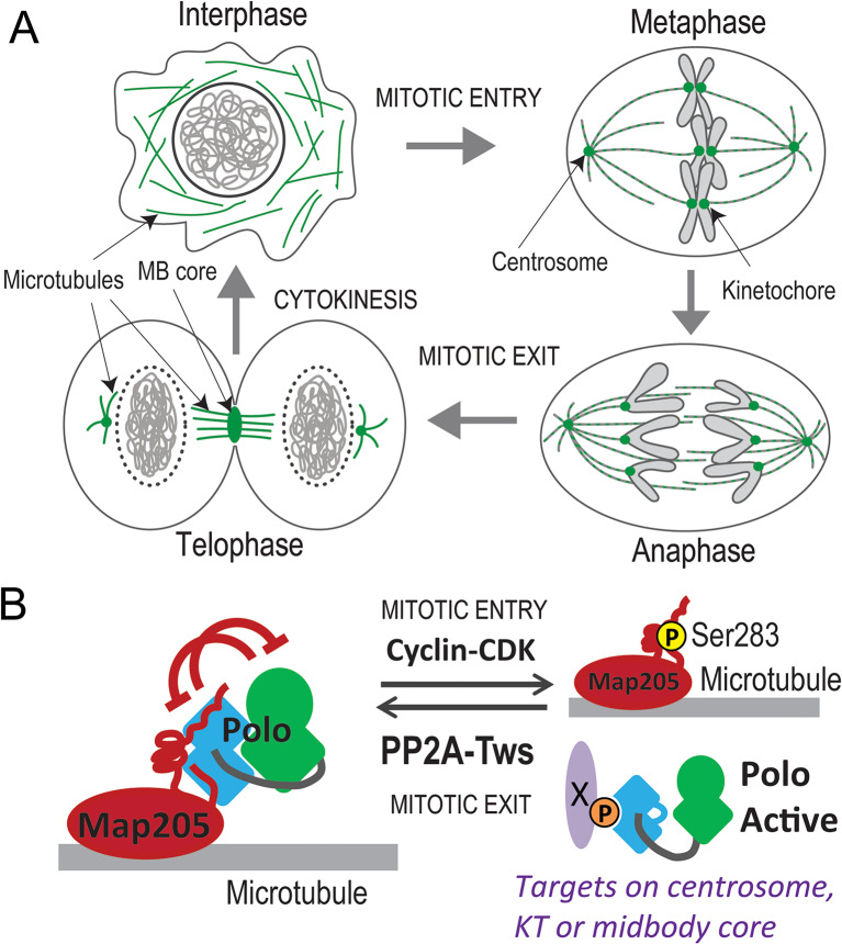
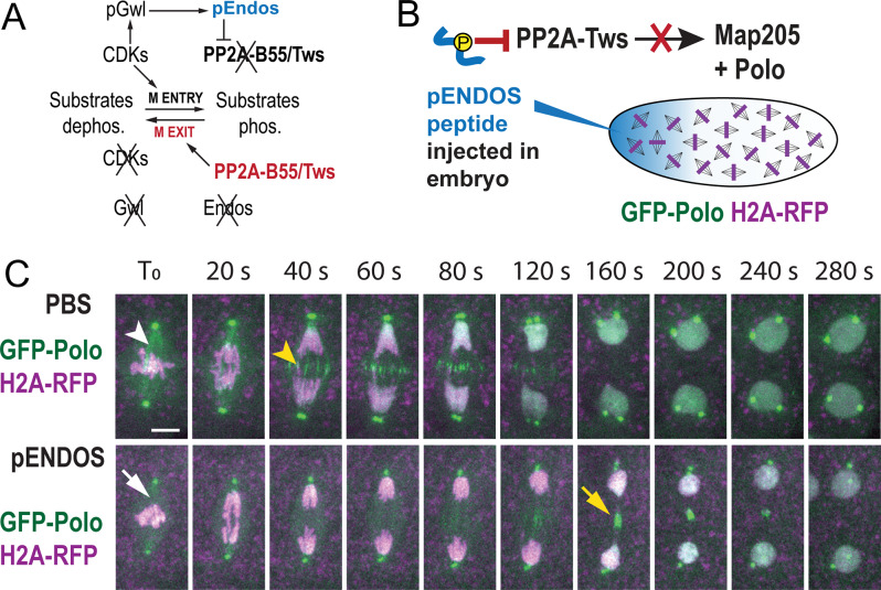
Background: Mitosis and cytokinesis are regulated by reversible phosphorylation events controlled by kinases and phosphatases. Drosophila Polo kinase, like its human ortholog PLK1, plays several roles in this process. Multiple mechanisms contribute to regulate Polo/PLK1 activity, localization and interactions. We previously showed that the microtubule-associated protein Map205 interacts with Polo during interphase and cytokinesis, inhibiting and sequestering Polo on microtubules. During mitosis, phosphorylation of Map205 at a Cyclin-Dependent Kinase site allows Polo to dissociate from Map205, when Polo must fulfill its mitotic functions. How the Polo/Map205 interaction is restored during mitotic exit remained unknown.

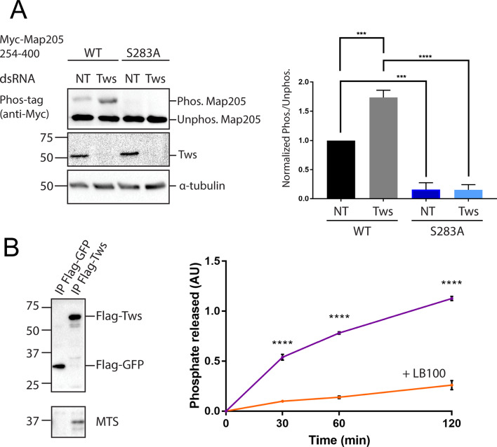
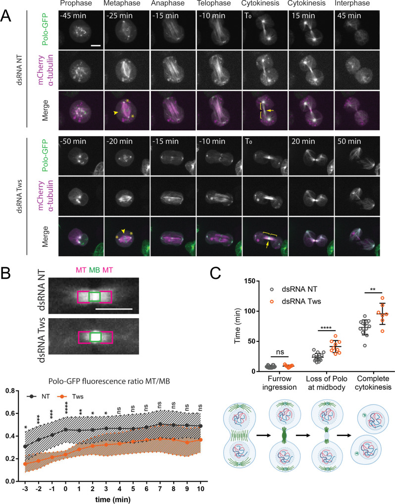
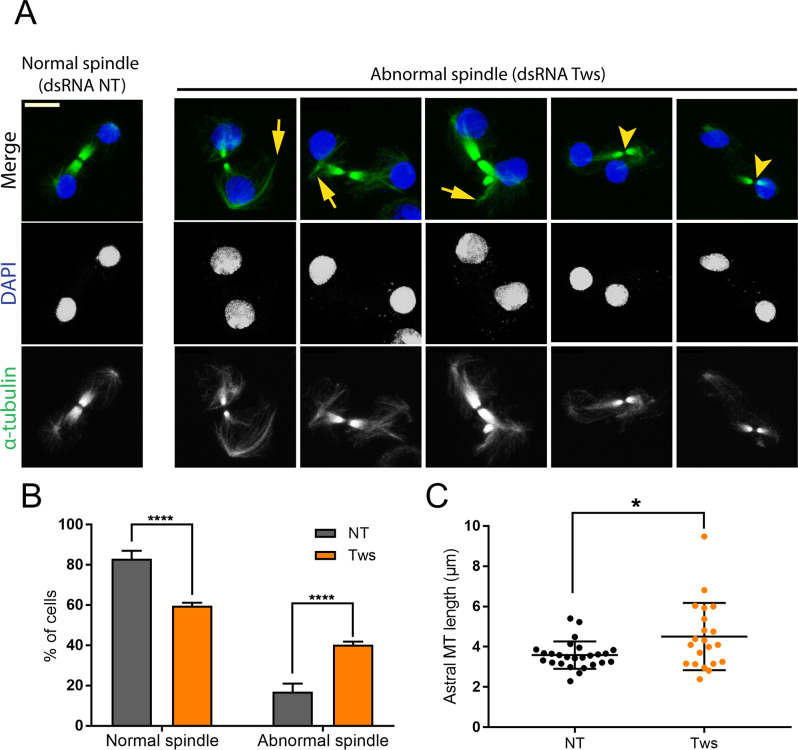
Results: Here we show that PP2A-Tws/B55 is required to dephosphorylate Map205, and enables the Map205-dependent localization of Polo to microtubules during cytokinesis. In addition, we show that PP2A-Tws is required for spindle function during cytokinesis, consistent with the essential role of Polo in this process.

Conclusions: These findings complement previous studies to provide an understanding of the full cycle of Polo regulation by Map205, kinases and phosphatases. Our findings have implications for the wider network of cell cycle regulatory circuitry.

Keywords: Drosophila; Cell cycle; Cytokinesis; Map205; Mitosis; PP2A-B55; Polo; Tws.


Conflict of interest statement

Declarations. Competing interests: The authors declare no competing interests.

Figures

Fig. 1
Fig. 1
Regulation of Polo localization and function during mitosis and cytokinesis. (A) The dynamic localization of Polo during the cell cycle. During mitosis (right), Polo (green) is strongly localized to kinetochores and centrosomes. During cytokinesis and interphase (left), Polo is strongly localized to microtubules. (B) Molecular regulation of Polo. During mitosis interphase (left), Polo interacts with Map205 on microtubules. This interaction stabilizes the PBD (blue) of Polo in a conformation that binds and inhibits the KD (green). During mitosis (right), mitotic Cyclin-CDK phosphorylation of Map205 causes the release of Polo. The phosphorylation of Polo interactors (X) facilitates their interactions with the PBD targeting Polo at centrosomes, kinetochores (KT) and the midbody core. During mitotic exit, Map205 dephosphorylation by PP2A-Tws promotes the recruitment of Polo by Map205 on microtubules
Fig. 2
Fig. 2
PP2A-Tws dephosphorylates Map205 at Ser283. (A) Map205 is hyperphosphorylated at Ser283 after depletion of RNAi depletion of Tws. Cells expressing Myc-Map205254 − 400 WT or S283A were transfected with dsRNA against Tws or Non-Target (NT). After 4 days, cells were analyzed by Western blotting using Phos-tag as indicated. Left: results from a representative experiment. Right: Normalized ratios of phosphorylated/unphosphorylated Myc-Map205254 − 400 band intensities. Averages of 3 experiments are shown. All error bars: S.D. *** p < 0.001, **** p < 0.0001 from paired t-tests. (B) PP2A-Tws dephosphorylates a Map205 peptide at Ser283 in vitro. Left: Flag-Tws or Flag-GFP were immunoprecipitated from cells and products were analyzed by Western blotting. Right: Dephosphorylation of the pSer283 Map205 peptide by Flag-Tws as a function of time (violet curve). Values obtained with Flag-GFP were subtracted. The Flag-Tws associated phosphatase activity is largely abrogated by the addition of LB100, a PP2A inhibitor (orange curve). Experiment repeated 3 times. Averages of technical triplicates from a representative experiment are shown. AU: arbitrary units. **** p < 0.0001 from unpaired t-tests
Fig. 3
Fig. 3
PP2A-Tws promotes the timely localization of Polo to microtubules and timely abscission during cytokinesis in D-Mel cells. (A) Cells expressing Polo-GFP and mCherry-Tubulin were filmed during their divisions. In control cells (RNAi NT, top), Polo-GFP becomes strongly localized to microtubules during telophase and cytokinesis. By contrast, in Tws-depleted cells (RNAi Tws, bottom), Polo-GFP is less strongly localized to microtubules (backets). However, the localization of Polo-GFP to centrosomes (asterisks), kinetochores (arrowheads) and the recently constricted midbody core (arrow) are not affected. Scale bar: 5 μm. (B) Quantification of Polo-GFP localization to microtubules relative to the midbody core during cytokinesis. Top: GFP fluorescence intensity measurements were taken on central spindle microtubules (MT, magenta boxes) and at the midbody core (MB, green boxes). Scale bar: 5 μm. Bottom: the ratio of MT/MB fluorescence over time was plotted as a function of time. T0: end of furrow ingression. Between 10 and 20 cells were quantified per condition. All error bars: S.D. * p < 0.05, **p < 0.01, ***<0.001, **** p < 0.0001, ns: non-significant from two-way Anova. (C) Quantifications of the durations of furrow ingression, retention of Polo-GFP at the midbody core and complete cytokinesis (until abscission) relative to anaphase onset in D-Mel cells depleted of Tws or control cells. The cartoons at the bottom illustrate the stages quantified. Between 7 and 13 cells were quantified in each condition. All error bars: S.D. * p < 0.05, **p < 0.01, ***<0.001, **** p < 0.0001, ns: non-significant from unpaired t-tests
Fig. 4
Fig. 4
PP2A-Tws promotes the localization of Polo to microtubules during karyokinesis in syncytial embryos. (A) PP2A-B55/Tws is inhibited by phosphorylated Endos during mitotic entry. During mitotic exit, PP2A-B55/Tws is reactivated when Endos is dephosphorylated. See text for more details. B-C. Injection of a phosphopeptide from Endos (pENDOS) to inhibit PP2A-Tws causes the mislocalization of GFP-Polo-GFP to the central spindle during karyokinesis. (B) Experimental scheme. (C) Transgenic embryos expressing GFP-Polo and H2A-RFP were imaged immediately after injection. T0: immediately before anaphase onset. After control injection (PBS only, top) during metaphase, GFP-Polo is faintly localized to microtubules already in metaphase (white arrowhead) and becomes concentrated on the central spindle after anaphase (yellow arrowhead). After injection of the pENDOS phospho-peptide, the localization of GFP-Polo to microtubules is strongly reduced (white arrow). In addition, GFP-Polo accumulates on the central spindle later, at the midbody core (yellow arrow). Scale bar: 5 μm
Fig. 5
Fig. 5
PP2A-Tws is required for spindle function during cytokinesis. (A) D-Mel cells were transfected with dsRNA against Tws or non-target (NT). After 3 days, cells were analyzed by immunofluorescence. The depletion of Tws results in cells with abnormal spindles showing bundles of microtubules that are distinct from or oriented away from the main spindle (arrows). Spindles are also mispositioned relative to daughter nuclei (arrowheads). (B) Quantification of abnormal vs. normal spindles from experiments as in A. Averages from 3 experiments are show. 100 cells were quantified per condition in each experiment. All error bars: S.D. **** p < 0.0001 from paired t-tests. (C) Quantification of maximal astral microtubule lengths from experiments as in A. Twenty-six (NT) and 21 (Tws) cells were quantified. * p = 0.02 from paired t-test

Similar articles

References

    1. Archambault V, Glover DM. Polo-like kinases: conservation and divergence in their functions and regulation. Nat Rev Mol Cell Biol. 2009;10:265–75. - PubMed
    1. Zitouni S, Nabais C, Jana SC, Guerrero A, Bettencourt-Dias M. Polo-like kinases: structural variations lead to multiple functions. Nat Rev Mol Cell Biol. 2014;15:433–52. - PubMed
    1. Petronczki M, Lenart P, Peters JM. Polo on the rise-from Mitotic Entry to Cytokinesis with Plk1. Dev Cell. 2008;14:646–59. - PubMed
    1. Sunkel CE, Glover DM. Polo, a mitotic mutant of Drosophila displaying abnormal spindle poles. J Cell Sci. 1988;89(Pt 1):25–38. - PubMed
    1. Archambault V, Lepine G, Kachaner D. Understanding the Polo kinase machine. Oncogene. 2015;34:4799–807. - PubMed

LinkOut - more resources