. 2024 Dec 28;24(1):435.
doi: 10.1186/s12935-024-03625-6.

Prognostic value and immune infiltration of a tumor microenvironment-related PTPN6 in metastatic melanoma

Affiliations

Prognostic value and immune infiltration of a tumor microenvironment-related PTPN6 in metastatic melanoma

Rongyao Sun et al. Cancer Cell Int. .

Abstract

Background: Cutaneous melanoma is one of the most invasive and lethal skin malignant tumors. Compared to primary melanoma, metastatic melanoma (MM) presents poorer treatment outcomes and a higher mortality rate. The tumor microenvironment (TME) plays a critical role in MM progression and immunotherapy resistance. This study focuses on the role of the TME-related gene PTPN6 in the prognosis and immunotherapy response of MM.

Methods: This study analyzed the RNA-seq and clinical data of MM patients from public databases, employing the ESTIMATE algorithm and bioinformatics tools to identify differentially expressed genes in the TME. PTPN6 was identified as a prognostic biomarker. Its expression and function were validated using in vitro and in vivo experiments. The role of PTPN6 in immune cell infiltration and its association with the JAK2-STAT3 pathway and immunotherapy response were also evaluated.

Results: PTPN6 expression was significantly lower in MM and associated with poor prognosis. In vitro, Overexpression of PTPN6 inhibited proliferation, migration, and invasion, while knockdown reversed these effects. In vivo, PTPN6 overexpression reduced tumor growth. Mechanistically, PTPN6 suppressed JAK2-STAT3 signaling pathway activation. High PTPN6 expression was positively associated with immune cell infiltration, improved immunotherapy response, and reduced PD-L1 expression.

Conclusion: The gene PTPN6, associated with the tumor microenvironment, may serve as a promising prognostic biomarker and therapeutic target for MM.

Keywords: Immunotherapy; JAK2-STAT3; Metastatic melanoma; PTPN6; Tumor microenvironment.


Conflict of interest statement

Declarations. Ethics approval and consent to participate: All animal experiments were approved by the Medical Ethics Committee of the Second Affiliated Hospital of Harbin Medical University (Ethical Review Approval Number: SYDW2024-074). Consent for publication: All authors gave their consent for publication. Competing interests: The authors declare no competing interests.

Figures
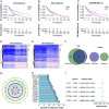
Fig. 1
Fig. 1
Identification of differentially expressed genes based on ImmuneScore and StromalScore. (A-C) Overall survival analysis of high and low Immune/Stromal/ESTIMATE Scores in patients with metastatic melanoma using the ESTIMATE algorithm. (D, E) Heatmap depicting the top 100 DEmRNAs with the most significant p-values between high and low Immune/Stromal Scores (|log Fold change|>1.9, p < 0.001). Blue represents downregulation, while red represents upregulation. (F) Venn diagram showing the upregulated and downregulated genes commonly intersecting between Stromal and Immune Scores, with green indicating immune-related DEmRNAs and purple indicating stromal-related DEmRNAs. (G) Construction of a protein-protein interaction network for overlapping genes using the STRING online tool (confidence score > 0.900) and Cytoscape software. (H) The top 15 genes ranked by nodes in the PPI network are shown in the graph. (I) Univariate Cox analysis was conducted on genes with a node count of 15 or more in the PPI network to identify differentially expressed genes significantly associated with prognosis
Fig. 2
Fig. 2
The expression of PTPN6 and its correlation analysis with survival. (A) Boxplot showing differential expression of PTPN6 in metastatic melanoma samples and normal skin tissue samples. (B) Correlation analysis of PTPN6 expression in metastatic melanoma samples with overall survival. (C) Correlation analysis of PTPN6 expression in metastatic melanoma samples with progression-free survival. (D) RT-qPCR assay analyzed the transcription level of PTPN6 in human keratinocyte cell line HaCaT and human metastatic melanoma cell lines A2058 and M14. The RT-qPCR results showed the relative mRNA expression of PTPN6 normalized to GAPDH. (E) Western blotting assay analyzed the expression of PTPN6 in human keratinocyte cell line HaCaT and human metastatic melanoma cell lines A2058 and M14. (**p < 0.01, ***p < 0.001)
Fig. 3
Fig. 3
Overexpression of PTPN6 inhibited the proliferation and viability of metastatic melanoma cells in vitro. (A–C) RT-qPCR and Western blotting assays were performed to evaluate the mRNA and protein expression of PTPN6 in A2058 and M14 cells after transfection with PTPN6. The RT-qPCR results showed the relative mRNA expression of PTPN6 normalized to GAPDH. (D, E) CCK8 assay was used to determine the impact of PTPN6 overexpression on the proliferation of A2058 and M14 cells. (F, G) EdU assay was conducted to assess the impact of PTPN6 overexpression on the viability of A2058 and M14 cells. Representative images were randomly selected from three independent experiments. Data are presented as the mean ± standard deviation (SD). (*p < 0.05, **p < 0.01, ***p < 0.001, ****p < 0.0001)
Fig. 4
Fig. 4
Overexpression of PTPN6 suppressed the migration and invasion of metastatic melanoma cells in vitro. (A, B) Wound healing assay was performed to determine the impact of PTPN6 overexpression on the migration of A2058 and M14 cells. (C, D) Transwell migration and Matrigel invasion assays were used to assess the impact of PTPN6 overexpression on the migration and invasion of A2058 and M14 cells. (E, F) Western blotting assay was conducted to verify the expression of migration and invasion-related genes, including MMP2, MMP9, E-cadherin, and Vimentin, in control, NC, and PTPN6 overexpression groups in A2058 and M14 cells. Representative images were randomly selected from three independent experiments. Data are presented as mean ± standard deviation (SD). (*p < 0.05, **p < 0.01, ***p < 0.001, ****p < 0.0001)
Fig. 5
Fig. 5
Overexpression of PTPN6 inhibited the activity of the JAK2-STAT3 signaling pathway in metastatic melanoma cells. (A-D) Western blotting assay was used to assess the expression of p-JAK2, JAK2, p-STAT3, and STAT3 after transfection with the PTPN6 overexpression plasmid. (****p < 0.0001)
Fig. 6
Fig. 6
PTPN6 may affect immune cell infiltration and immune therapy response in metastatic melanoma. (A) TIMER 2.0 online analysis tool was used to examine the relationship between PTPN6 and the levels of infiltration of 14 immune cells and tumor purity in metastatic melanoma. (B) The correlation between PTPN6 and immune checkpoint therapy (alone or in combination with PD-1 or CTLA-4) response was analyzed using the TCIA database. (C, D) Western blotting assay was used to detect PD-L1 expression after PTPN6 overexpression (*p < 0.05, **p < 0.01)
Fig. 7
Fig. 7
Knockdown of PTPN6 promoted the proliferation, migration, and invasion of metastatic melanoma cells in vitro. (A, B) Western blotting assay was performed to evaluate the protein expression of PTPN6 in A2058 and M14 cells after transfection with GV341-PTPN6-3FLAG-SV40-puromycin lentivirus. (C, D) Western blotting assay was performed to evaluate the protein expression of PTPN6 in A2058 and M14 cell lines stably overexpressing PTPN6 after transiently transfected siPTPN6 or siNC. (E, F) CCK8 assay was used to determine the impact of PTPN6 knockdown on the proliferation of cells. (G, H) Wound healing assay was performed to determine the impact of PTPN6 knockdown on the migration of cells. (I, J) Transwell migration and Matrigel invasion assays were used to assess the impact of PTPN6 knockdown on the migration and invasion of cells. Representative images were randomly selected from three independent experiments. Data are presented as mean ± standard deviation (SD). (**p < 0.01, ***p < 0.001, ****p < 0.0001)
Fig. 8
Fig. 8
Overexpression of PTPN6 inhibited the growth of MM cells in vivo. (A-D) BALB/c nude mice were injected subcutaneously with control and stably PTPN6 overexpressing A2058 or M14 cells. (E, F) The tumor volume growth curves of mice were plotted. (G, H) Measurement of mice tumor weight. (I, J) IHC analysis of PTPN6, Ki67 and MMP9 expressions in tumor tissues. Representative images were randomly selected from three independent experiments. Data are presented as mean ± standard deviation (SD). (*p < 0.05, **p < 0.01, ***p < 0.001, ****p < 0.0001)

Similar articles

Cited by

References

    1. Garbe C, Amaral T, Peris K, Hauschild A, Arenberger P, Basset-Seguin N, et al. European consensus-based interdisciplinary guideline for melanoma. Part 1: Diagnostics: Update 2022. Eur J Cancer. 2022;170:236–55. - PubMed
    1. Long GV, Swetter SM, Menzies AM, Gershenwald JE, Scolyer RA. Cutaneous melanoma. Lancet. 2023;402(10400):485–502. - PubMed
    1. Garbe C, Amaral T, Peris K, Hauschild A, Arenberger P, Basset-Seguin N, et al. European consensus-based interdisciplinary guideline for melanoma. Part 2: treatment - update 2022. Eur J Cancer. 2022;170:256–84. - PubMed
    1. Davis LE, Shalin SC, Tackett AJ. Current state of melanoma diagnosis and treatment. Cancer Biol Ther. 2019;20(11):1366–79. - PMC - PubMed
    1. Eddy K, Chen S. Overcoming Immune Evasion in Melanoma. Int J Mol Sci. 2020;21(23):8984. - PMC - PubMed

LinkOut - more resources