. 2024 Dec 28;13(1):123.
doi: 10.1186/s40164-024-00586-4.

AOH1996 targets mitochondrial dynamics and metabolism in leukemic stem cells via mitochondrial PCNA inhibition

Affiliations

AOH1996 targets mitochondrial dynamics and metabolism in leukemic stem cells via mitochondrial PCNA inhibition

HyunJun Kang et al. Exp Hematol Oncol. .

Erratum in

Abstract

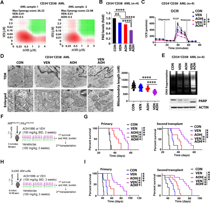
Cytoplasmic proliferating cell nuclear antigen (PCNA) is highly expressed in acute myeloid leukemia (AML) cells, supporting oxidative metabolism and leukemia stem cell (LSC) growth. We report on AOH1996 (AOH), an oral compound targeting cancer-associated PCNA, which shows significant antileukemic activity. AOH inhibited growth in AML cell lines and primary CD34 + CD38 - blasts (LSC-enriched) in vitro while sparing normal hematopoietic stem cells (HSCs). In vivo, AOH-treated mice demonstrated prolonged survival compared to controls (50 vs. 35 days; p < 0.0001) with reduced LSC burden, as shown in secondary transplants (42 vs. 30 days, p < 0.0001). Mechanistically, AOH disrupted mitochondrial PCNA's binding to the OPA1 protein, enhancing OPA1's interaction with its E3 ligase, MARCH5, which led to OPA1 degradation. This process reduced mitochondrial length, fatty acid oxidation (FAO), and oxidative phosphorylation (OXPHOS), thereby inhibiting LSC expansion. The addition of venetoclax (VEN), an FDA-approved Bcl-2 inhibitor, further enhanced AOH's effects, reducing mitochondrial length, FAO, and OXPHOS while improving survival in AML models. While VEN is approved for AML, AOH is under clinical investigation for solid tumors, and our findings support its broader therapeutic potential.

Keywords: AML; AOH1996; Leukemic stem cells; Mitochondrial metabolism; PCNA inhibitor.


Conflict of interest statement

Declarations. Ethics approval and consent to participate: Healthy donor-derived normal hematopoietic stem cells (HSCs) and acute myeloid leukemia (AML) specimens were sourced from the City of Hope National Medical Center (COHNMC) in accordance with approved banking protocols (#06229, #03162, #07047, or #18067) sanctioned by the City of Hope Institutional Review Board. These protocols adhere to the guidelines set forth by the Department of Health and Human Services and are compliant with the principles outlined in the Declaration of Helsinki. Prior to specimen acquisition, written informed consent was obtained from donors (#06229) or patients (#03162, #07047, or #18067). Consent for publication: Not applicable. Competing interests: The authors declare no competing interests.

Figures

Fig. 1
Fig. 1
Impact of AOH1996 (AOH) on leukemic stem cells and leukemogenesis via inhibition of mitofusion and mitochondrial metabolism. A Effects of AOH (1 µM) on proliferation and apoptosis of LSC-enriched AML blasts. CD34 + CD38- cells were isolated from primary MNCs (n = 4) or AML blasts (n = 4). Top, cell proliferation levels. Bottom, apoptosis levels. B Effects of AOH on colony formation of LSC-enriched AML blasts. CD34 + CD38- AML blasts (top) or MNCs (bottom) (2 × 10⁵ cells/mL, n = 3) were treated with DMSO control (CON) or indicated doses of AOH for 24 h, then plated in methylcellulose. After 14 days, colonies were imaged using a light microscope and counted. Data are shown as mean ± SE, with triplicate determinations. C Unsupervised hierarchical clustering of significantly different (adj. p < 0.05) metabolites from primary CD34 + AML blasts treated with AOH (0.5 µM), DMSO or Non-treat controls for 24 h. Metabolite changes were displayed as a heat map. D-F Primary CD34 + CD38- AML blasts were treated with VEH or AOH (1 µM) for 24 h. D Effects of AOH on FAO (measured by ³H-palmitate levels, top) and OXPHOS (indicated by OCR levels, bottom), with the OCR comparison focused on maximal respiratory capacity. E Transmission electron microscopy (TEM) imaging of mitochondria. Enlarged images are shown. Scale bar, 1 μm. F Quantification of mitochondrial length (n = 30). Asterisks indicate statistically significant differences based on unpaired t-test analysis. G-J AOH effects on mitochondrial PCNA’s interaction with OPA1 and its impact on OPA1 stability in CD34 + CD38- AML blasts treated with VEH or AOH (1 µM) for 24 h. G Immunoblot analysis of mitofusion-regulated and mitochondrial metabolism-regulated proteins. H Mitochondrial fractions from treated cells were immunoprecipitated with anti-PCNA and immunoblotted with anti-OPA1 antibodies. Input loading controls are shown. I Cells were treated with cycloheximide, a translation inhibitor, (CHX, 10 µM), to assess protein stability, for indicated times, and lysates were immunoblotted with anti-OPA1 antibodies. J Left, lysates were immunoprecipitated with anti-OPA1 and immunoblotted with anti-MARCH5 antibodies. Input loading controls are shown. Right, ubiquitination assay with lysates immunoprecipitated with anti-OPA1 and immunoblotted with anti-Ub antibodies. K-L Antileukemic activities of AOH in vivo. Human primary AML blasts (1.0 × 10⁶) were injected intravenously into Es1(ko) SCID mice. After 7 days, mice were treated with vehicle control or AOH (100 mg/kg, BID, oral gavage, 3 weeks). K Leukemia burden at day 17 post-transplant, measured by the percentage of human CD45 + cells (left) and spleen size (right). L Left, effects of AOH treatment in vivo on FAO/OXPHOS in human CD45 + cells isolated from treated mice. FAO (top) and OXPHOS (bottom, indicated by OCR) levels were measured. Right, top Kaplan–Meier survival curve of primary transplanted leukemic mice treated with CON (black line, n = 10) and AOH-treated mice (red line, n = 10). Median survival (MS): 35 days (CON), 50 days (AOH). Right, bottom, Kaplan–Meier survival curve of secondary transplanted leukemic mice treated with CON (black line, n = 10) and AOH-treated mice (red line, n = 10). MS: 30 days (CON), 42 days (AOH). Statistical significance determined by Log-rank (Mantel–Cox) test (p < 0.0001)
Fig. 2
Fig. 2
Synergistic effects of AOH and VEN in vitro and in vivo. A Synergistic effect of AOH and VEN on LSC-enriched AML blasts. Two primary CD34 + CD38- AML samples were used: AML sample-1 (corresponding to AML-2 in Table S2) and AML-sample 2 (corresponding to AML-3 in Table S2). Cells (1 × 105 cells/mL, n = 3) were treated with indicated concentration of AOH and VEN. Levels of cell proliferation were evaluated and synergy score of the drug combination was calculated. Maximum synergy scores were 26.32 for AML sample-1 and 22.98 for AML sample-2. B-D Combinatorial effects of AOH and VEN on FAO/OXPHOS levels and mitochondrial length of LSC-enriched AML blasts. Primary CD34 + CD38- AML blasts (n = 4) were treated with DMSO (CON), AOH (1 µM), VEN (20 nM), or combination of AOH and VEN for 24 h. B FAO levels. C OXPHOS levels (indicated by OCR), with OCR comparisons focused on maximal respiratory capacity. D Mitochondria length. Left, represented TEM images. Right, quantification of mitochondria length (n = 30). E Primary CD34 + CD38- AML blasts (n = 4) were treated with DMSO (CON), VEN (20 nM), AOH (1 µM), or combination of AOH and VEN for 24 h. Top, DNA fragmentation. Bottom, PARP cleavage. ANOVA test was performed for multiple group comparisons prior to statistical analysis of each two group comparisons. F-G Combinatorial effects of AOH and VEN on MllPTD/WT/Flt3ITD/ITD AML mouse model. Kaplan-Meier curve employing log-rank test was used to find statistical significance. F Experimental design for AOH and VEN combined treatment. 1 × 106 MllPTD/WT/Flt3ITD/ITD BM MNCs were intravenously injected into normal Ces1c(ko) B6 WT recipients. The transplanted mice were then randomly divided into 4 groups 7 days post-transplant (n = 10/group) and treated with either vehicle (CON), AOH (100 mg/kg, BID, PO, 21 days), VEN (100 mg/kg, daily, PO, 21 days) or AOH/VEN at the same doses of single agents. On day 28 post-transplant, 106 BM MNCs cells from each treatment group were harvested for secondary transplant. G Left, Kaplan–Meier survival curve of primary transplanted leukemic mice treated with CON (black line, MS 34 days), VEN (blue line, MS 34 days), AOH (red line, MS 43.5 days), or AOH/VEN (purple line, MS 54 days). Right, Kaplan–Meier survival curve of secondary transplanted leukemic mice treated with CON (black line, MS 28 days), VEN (blue line, MS 32.5 days), AOH (red line, MS 41 days), or AOH/VEN (purple line, MS 49.5 days). H-I Combinatorial effects of AOH and VEN on FLT3-WT PDX AML model. H Experimental design for AOH and VEN combined treatment. hCD45 + BM FLT3-WT AML cells (1 × 106 cells/mouse) were transplanted into Es1(ko) SCID mice to generate a cohort of AML bearing PDX mice. The transplanted mice were then randomly divided into 4 groups 7 days post-transplant (n = 10/group) and treated with either vehicle (CON), AOH (100 mg/kg, BID, PO, 21 days), VEN (100 mg/kg, daily, PO, 21 days) or AOH/VEN at the same doses of single agents. On day 28, 106 BM MNCs cells from each treatment group were harvested for secondary transplant. I Left, Kaplan–Meier survival curve of primary transplanted leukemic mice treated with CON [black line, median survival (MS) 41 days], VEN (blue line, MS 48 days), AOH (red line, MS 55 days), or AOH/VEN (purple line, MS 75.5 days). Right, Kaplan–Meier survival curve of secondary transplanted leukemic mice treated with CON (black line, MS 40 days), VEN (blue line, MS 51 days), AOH (red line, MS 60 days), or AOH/VEN (purple line, MS 76 days)

References

    1. Strzalka W, Ziemienowicz A. Proliferating cell nuclear antigen (PCNA): a key factor in DNA replication and cell cycle regulation. Ann Bot. 2011;107(7):1127–40. - PMC - PubMed
    1. Wang L, Kong W, Liu B, Zhang X. Proliferating cell nuclear antigen promotes cell proliferation and tumorigenesis by up-regulating STAT3 in non-small cell lung cancer. Biomed Pharmacother. 2018;104:595–602. - PubMed
    1. Naryzhny SN, Lee H. Proliferating cell nuclear antigen in the cytoplasm interacts with components of glycolysis and cancer. FEBS Lett. 2010;584(20):4292–8. - PubMed
    1. Røst LM, Ræder SB, Olaisen C, et al. PCNA regulates primary metabolism by scaffolding metabolic enzymes. Oncogene. 2023;42(8):613–24. - PMC - PubMed
    1. Ohayon D, De Chiara A, Chapuis N, et al. Cytoplasmic proliferating cell nuclear antigen connects glycolysis and cell survival in acute myeloid leukemia. Sci Rep. 2016;6(1):35561. - PMC - PubMed

LinkOut - more resources