. 2024 Dec 28;14(1):31291.
doi: 10.1038/s41598-024-82614-1.

Curcumin liposomes alleviate senescence of bone marrow mesenchymal stem cells by activating mitophagy

Affiliations

Curcumin liposomes alleviate senescence of bone marrow mesenchymal stem cells by activating mitophagy

Weiyao Li et al. Sci Rep. .

Abstract

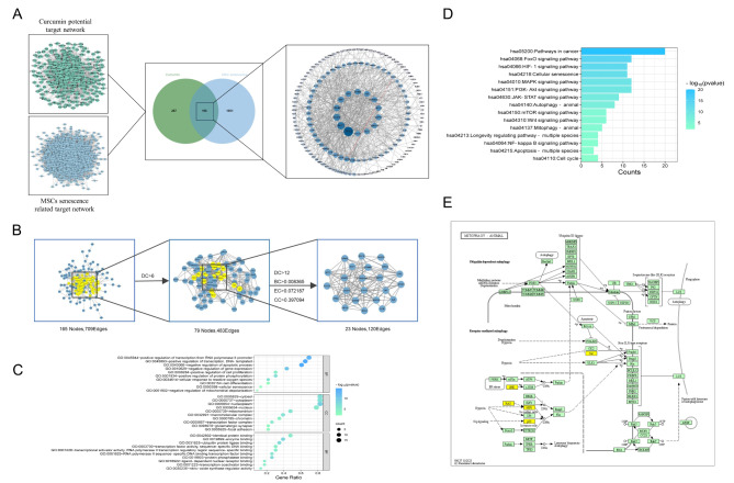
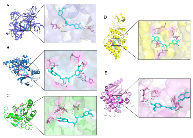
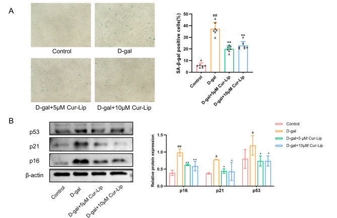
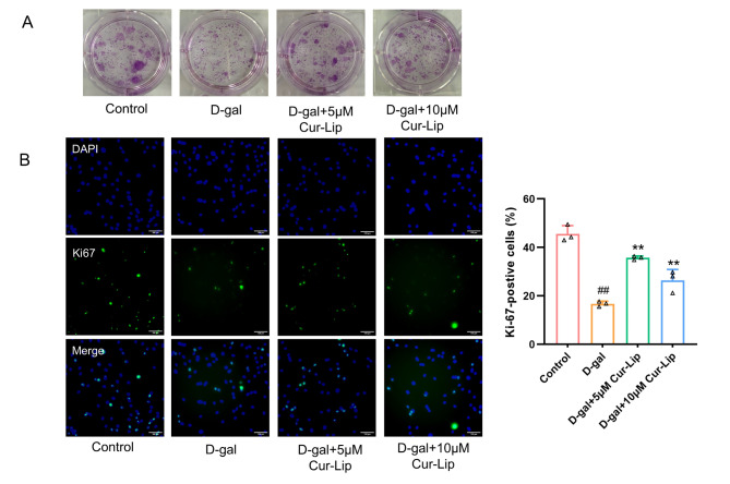
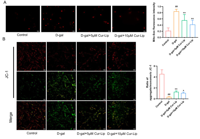
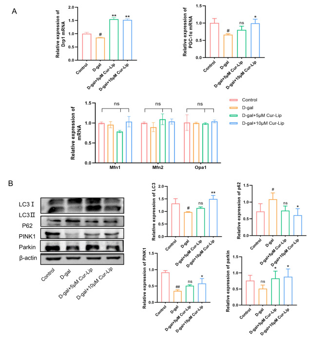
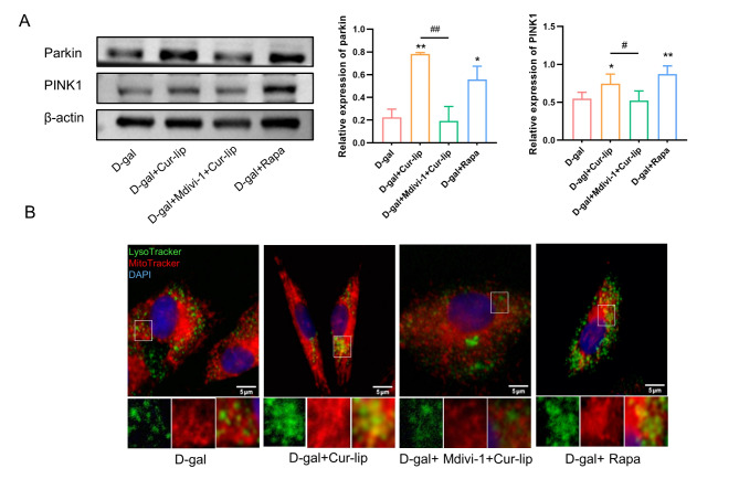
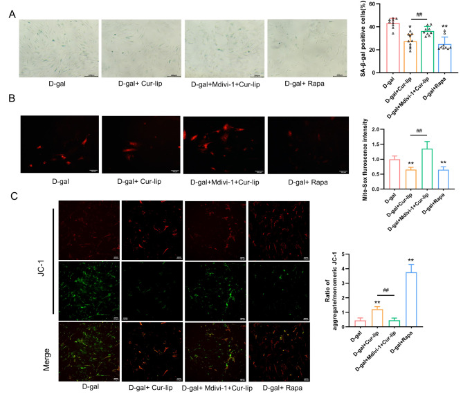
The senescence of mesenchymal stem cells (MSCs) is closely related to aging and degenerative diseases. Curcumin exhibits antioxidant and anti-inflammatory effects and has been extensively used in anti-cancer and anti-aging applications. Studies have shown that curcumin can promote osteogenic differentiation, autophagy and proliferation of MSCs. Liposome, as a nano-carrier, provides a feasible strategy for improving the bioavailability and controlled-release profile of curcumin.This study aimed to evaluate the effects of curcumin liposomes (Cur-Lip) on the senescence of rat bone marrow mesenchymal stem cells (rBMSCs). Based on network pharmacology, we predicted the targets and mechanisms of curcumin on senescence of MSC. 23 key targets of Cur were associated with MSC senescence were screened out and mitophagy signaling was significantly enriched. Cur-Lip treatment alleviated senescence of D-galactose (D-gal)-induced rBMSCs, protected mitochondrial function, and activated mitophagy, which may be related to mitochondrial fission. Inhibition of mitophagy attenuated the protective effects of Cur-lip on mitochondrial function and senescence of rBMSCs. Our findings suggested that Cur-Lip could alleviate senescence of rBMSC and improve mitochondrial function by activating mitophagy.

Keywords: Curcumin liposomes; Mitophagy; Network pharmacology; Senescence; rBMSCs.


Conflict of interest statement

Declarations. Competing interests: The authors declare no competing interests. Ethical approval: The study was approved by the Ethics Committee of the institutional Animal Care and Use Committee of Southwest Medical University (Approval No. swmu20230085).

Figures

Fig. 1
Fig. 1
Network pharmacology analysis predicted the mechanisms of the Cur effect on the senescence of MSCs. (A) The screening process for Cur-senescent MSCs co-targets. (B) Topology analysis and key target screening. (C) GO enrichment analysis of BP, CC, and MF. (D) KEGG enrichment analysis of anti MSCs senescence core target of Cur. (E) Mitophagy pathway.
Fig. 2
Fig. 2
Molecular docking between Cur and mitophagy-related proteins. (A) Cur-TP53. (B) Cur-MAPK8. (C) Cur-HRAS. (D) Cur-SRC. (E) Cur-HIF1α. Dotted line: molecular binding site.
Fig. 3
Fig. 3
Cur-Lip alleviated D-gal induced senescence of rBMSCs. (A) SA-β-gal assay in rBMSCs. Scale bar = 150 μm. (B) The effects of Cur-Lip on the protein expression of p16, p21 and p53 in rBMSCs. *p < 0.05, ** p < 0.01 vs. D-gal group; # p < 0.05, ## p < 0.01 vs. control group (same as below).
Fig. 4
Fig. 4
Cur-Lip improved cell proliferation in senescent rBMSCs. (A) Clone formation assay. (B) Representative images of Ki67 staining in rBMSCs. Scale bar = 100 μm.
Fig. 5
Fig. 5
The protective effect of Cur-Lip on mitochondrial function in rBMSCs. (A) Mitochondrial ROS generation. Scale bar = 100 μm. (B) Mitochondrial membrane potential. Scale bar = 100 μm.
Fig. 6
Fig. 6
Cur-Lip promoted mitochondrial quality control in rBMSCs. (A) The mRNA expression of Opa1, Mfn1, Mfn2, Drp1 and PGC-1α in rBMSCs. (B) Cur-Lip increased the expression levels of mitophagy-related proteins in rBMSCs.
Fig. 7
Fig. 7
The level of mitophagy after treatment with each group. (A) Expression levels of mitophagy-specific proteins PINK1 and Parkin. (B) Co-localization assay in rBMSCs with Lyso-Tracker (green) and Mito-Tracker (Red). Blue staining = DAPI. Scale bars = 5 μm. *p < 0.05, ** p < 0.01 vs. D-gal group; #p < 0.05, ##p < 0.01vs Cur-Lip group (same as below).
Fig. 8
Fig. 8
Mitophagy inhibited the senescence of rBMSCs. (A) SA-β-gal assay in rBMSCs. Scale bars = 200 μm. (B) Mitochondrial ROS generation. Scale bars = 100 μm. (C) Mitochondrial membrane potential.

Similar articles

Cited by

References

    1. Fitzsimmons, R., Mazurek, M. S., Soos, A. & Simmons, C. A. Mesenchymal stromal/stem cells in regenerative medicine and tissue engineering. Stem Cells Int.2018, 8031718–8031733 (2018). - PMC - PubMed
    1. Zhu, Y., Ge, J., Huang, C., Liu, H. & Jiang, H. Application of mesenchymal stem cell therapy for aging Frailty: From mechanisms to therapeutics. Theranostics11, 5675–5685 (2021). - PMC - PubMed
    1. Chen, H., Liu, O., Chen, S. & Zhou, Y. Aging and mesenchymal stem cells: Therapeutic opportunities and challenges in the Older Group. Gerontology68, 339–352 (2022). - PMC - PubMed
    1. Abruzzo, P. M. et al. Herb-derived products: Natural tools to delay and counteract stem cell senescence. Stem Cells Int.2020, 8827038–8827064 (2020). - PMC - PubMed
    1. Vono, R., Jover, G. E., Spinetti, G. & Madeddu, P. Oxidative stress in mesenchymal stem cell senescence: Regulation by coding and noncoding Rnas. Antioxid. Redox Signal.29, 864–879 (2018). - PMC - PubMed

Publication types

LinkOut - more resources