. 2024 Dec 28;14(1):31431.
doi: 10.1038/s41598-024-83024-z.

Development of chimeric MrNV virus-like particles capable of binding to SARS-CoV-2-susceptible cells and reducing infection by pseudovirus variants

Affiliations

Development of chimeric MrNV virus-like particles capable of binding to SARS-CoV-2-susceptible cells and reducing infection by pseudovirus variants

Supawich Boonkua et al. Sci Rep. .

Abstract

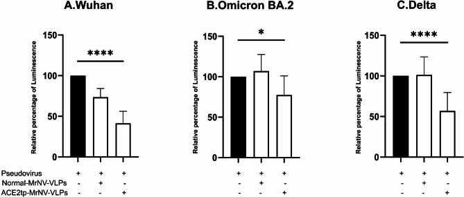
SARS-CoV-2, the cause of COVID-19, primarily targets lung tissue, leading to pneumonia and lung injury. The spike protein of this virus binds to the common receptor on susceptible tissues and cells called the angiotensin-converting enzyme-2 (ACE2) of the angiotensin (ANG) system. In this study, we produced chimeric Macrobrachium rosenbergii nodavirus virus-like particles, presenting a short peptide ligand (ACE2tp), based on angiotensin-II (ANG II), on their outer surfaces to allow them to specifically bind to ACE2-overexpressing cells called ACE2tp-MrNV-VLPs. Replacing the ACE2tp at the protruding domains (P-domain) of the MrNV capsid proteins did not affect their normal assembly into icosahedral VLPs. The presentation of the ACE2tp on the P-domains significantly improved the binding and internalization of ACE2tp-MrNV-VLPs to hACE2-overexpressing HEK293T cells in a concentration-dependent manner. Furthermore, ACE2tp-MrNV-VLPs exhibited the ability to block the binding and infection of SARS-CoV-2 pseudovirus variants, including Wuhan, BA.2 Omicron, and Delta subtypes. Our results suggest that chimeric ACE2tp-MrNV-VLPs can serve as a blocking agent against various SARS-CoV-2 mutated variants and could also potentially serve as target-specific nano-containers to carry therapeutic agents to combat SARS-CoV-2 infections in the future.

Keywords: Macrobrachium rosenbergii nodavirus (MrNV); Genetic modification; Nanotechnology; Recombinant capsid protein; SARS-CoV-2; Virus-like particle.


Conflict of interest statement

Declarations. Competing interests: The authors declare no competing interests.

Figures

Fig. 1
Fig. 1
ACE2tp-MrNV-capsid protein layout and structural prediction. (A) The schematic diagram of the layout of the normal MrNV and chimeric ACE2tp-MrNV-capsid proteins, with the ACE2tp peptide replacement shown in red. (B) depicts the ribbon diagram of the normal MrNV-VLP capsid protein (leftmost panel, P-domain = protruding domain, S-domain = shell domain). It also shows an icosahedral VLP modeling of the 180 capsid protein subunits from the 5-fold view, including a detailed look at the protruding domains (right panel with inset). (C) shows the ribbon diagram of the ACE2tp-MrNV-VLP capsid protein (leftmost panel, red loop indicating the ACE2tp peptide). Additionally, an icosahedral modeling of the ACE2tp-MrNV-VLP is provided, featuring a detailed view of the protruding domains, highlighting the ACE2tp peptide in red (right panel with inset).
Fig. 2
Fig. 2
Expression, purification, and assembly of ACE2tp-MrNV-capsid proteins into icosahedral virus-like particles (VLPs). (A) SDS-PAGE with R250 Coomassie Blue staining and Western blot using an anti-6xHisTag revealed protein bands corresponding to the molecular weights (MW) of approximately 42.5 kDa for normal MrNV-VLPs and approximately 43 kDa for ACE2tp-MrNV capsid proteins (N = normal MrNV-VLP, A = ACE2tp-MrNV-VLP). Electron micrographs of normal MrNV-VLPs (B, upper panel) and ACE2tp-MrNV-VLPs (C, lower panel) as displayed by negative staining. The average size of all VLP particles formed was in the range of 26–28 nm. Scale bars = 100 nm.
Fig. 3
Fig. 3
Binding of ACE2tp-MrNV-VLPs to HEK293T-hACE2 protein lysates and cells. (A) shows the levels of normal MrNV-VLPs and ACE2tp-MrNV-VLP binding to HEK239T cell lysates.(B) shows the levels of normal MrNV-VLPs and ACE2tp-MrNV-VLP binding to ACE2-overexpressing cell lysates. A and B were quantified using ELISA. The VLPs were incubated with the cell lysate-coated well at dilutions ranging from 0.3125, 0.625, 1.25, 2.5 and 5 µg. Data are presented as mean ± SD. (C) shows HEK293T cells incubated with either normal MrNV and ACE2tp-MrNV-VLPs. (D) shows HEK293T-hACE2 cells incubated with either normal MrNV or ACE2tp-MrNV-VLPs. (C) and (D) were stained with anti-6xHisTag antibody, and the corresponding secondary antibody conjugated with Alexa-594 (red fluorescence) and observed by confocal microscopy. Scale bars = 40 μm.
Fig. 4
Fig. 4
Internalization of ACE2tp-MrNV-VLPs into HEK293T-hACE2 cells. Chimeric ACE2tp-MrNV-VLPs (lower panels) showed a significant increase in internalization (red fluorescence) into HEK293T-hACE2 cells (cell nuclei observable as blue fluorescence) compared to control (upper panels). Scale bars = 40 μm.
Fig. 5
Fig. 5
Effect of ACE2tp-MrNV-VLPs on the level of infection by SARS-CoV-2 S-protein pseudovirus variants in HEK293T-hACE2 cells. Wuhan (A), Omicron BA.2 (B), and Delta (C) pseudovirus subtypes were used to infect the cells (column 1), compared to cells that were pre-incubated with normal MrNV-VLPs (column 2) and ACE2tp-MrNV-VLPs (column 3). Luminescence levels indicating infection in the HEK293T-hACE2 cells were measured after 48 h. (* p ≤ 0.05, ** p ≤ 0.01, *** p ≤ 0.001 **** p < 0.0001).
Fig. 6
Fig. 6
Schematic illustrations of the strategies to reduce SARS-CoV-2 interaction with ACE2 receptor. (A) Mutations and mode of interaction of SARS-CoV-2 variants with the ACE2 receptor. (B) The strategy of blocking SARS-CoV-2 virus entry by ACE2tp-MrNV-VLPs. (C) Future strategy for ACE2tp-MrNV-VLPs encapsulation with anti-viral agents against SARS-CoV-2.

Similar articles

Cited by

References

    1. Huang, C. et al. Clinical features of patients infected with 2019 novel coronavirus in Wuhan, China. Lancet395 (10223), 497–506 (2020). - PMC - PubMed
    1. Lu, R. et al. Genomic characterisation and epidemiology of 2019 novel coronavirus: Implications for virus origins and receptor binding. Lancet395 (10224), 565–574 (2020). - PMC - PubMed
    1. Magazine, N. et al. Mutations and evolution of the SARS-CoV-2 spike protein. Viruses14(3), 640 (2022). - PMC - PubMed
    1. Chavda, V. P. et al. mRNA-Based vaccines and therapeutics for COVID-19 and future pandemics. Vaccines (Basel)10(12), 2150 (2022) - PMC - PubMed
    1. Spenlehauer, C., Kirn, A., Aubertin, A. M. & Moog, C. Antibody-mediated neutralization of primary human immunodeficiency virus type 1 isolates: Investigation of the mechanism of inhibition. J. Virol.75 (5), 2235–2245 (2001). - PMC - PubMed

Publication types

MeSH terms

Supplementary concepts