. 2024 Dec 28;14(1):31338.
doi: 10.1038/s41598-024-82814-9.

S100A4 contributes to colorectal carcinoma aggressive behavior and to chemoradiotherapy resistance in locally advanced rectal carcinoma

Affiliations

S100A4 contributes to colorectal carcinoma aggressive behavior and to chemoradiotherapy resistance in locally advanced rectal carcinoma

Yohei Harada et al. Sci Rep. .

Abstract

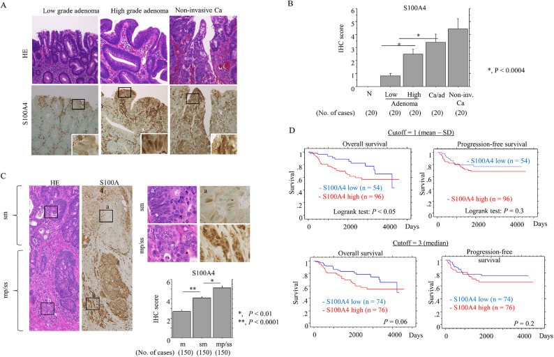
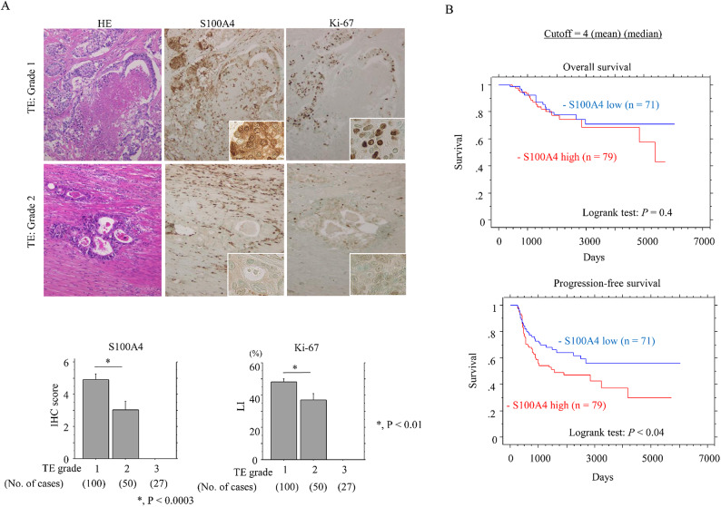
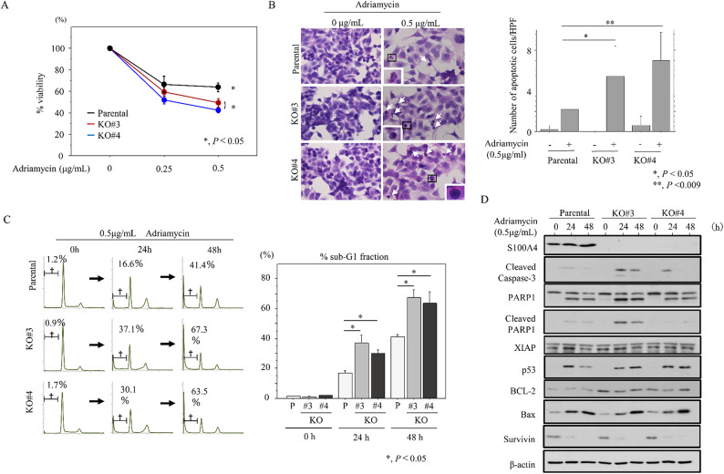
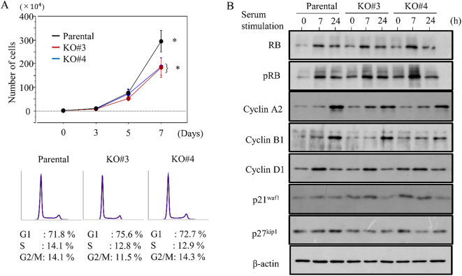
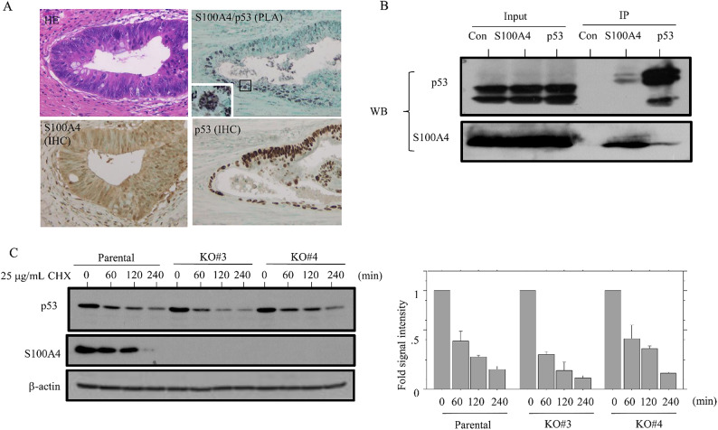
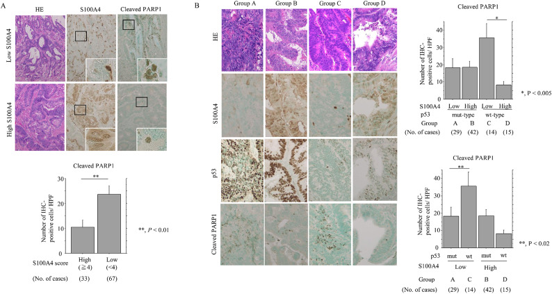
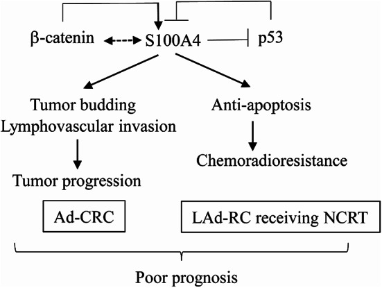
To investigate the functional role of S100A4 in advanced colorectal carcinoma (Ad-CRC) and locally advanced rectal carcinoma (LAd-RC) receiving neoadjuvant chemoradiotherapy (NCRT). We analyzed histopathological and immunohistochemical sections from 150 patients with Ad-CRC and 177 LAd-RC patients treated with NCRT. S100A4 knockout (KO) HCT116 cells were also used. S100A4 expression was absent in normal mucosa but increased progressively from colorectal adenoma to carcinoma, suggesting that S100A4 regulation is an early event in colorectal carcinogenesis. In Ad-CRC, high S100A4 expression correlated with high tumor budding and nuclear β-catenin, deep invasion, lymph-vascular involvement, and unfavorable prognosis. In NCRT-treated LAd-RC, high S100A4 expression was associated with poor treatment response and short progression-free survival. S100A4 KO decreased the proliferation of HCT116 cells through activation of the p53/p21waf1 axis, and sensitized cells to adriamycin-induced apoptosis. Levels of the apoptotic marker, cleaved poly (ADP-ribose) polymerase 1, were significantly higher in samples with low S100A4 and wild type p53. Finally, we observed a direct interaction between S100A4 and p53. In conclusion, S100A4 expression engenders aggressive behavior in Ad-CRC through association with β-catenin-driven tumor buddings. S100A4 exerts anti-apoptotic and proliferative effects via inhibition of p53 in LAd-RC patients receiving NCRT, which leads to chemoradioresistance and poor prognosis.

Keywords: Apoptosis; Colorectal carcinoma; Neoadjuvant chemoradiotherapy; S100A4; p53.


Conflict of interest statement

Declarations. Competing interests: The authors declare no competing interests. Consent for publication: Not applicable. Ethics approval: This study was conducted in accordance with the principles expressed in the Declaration of Helsinki 1964 and later versions. This study was approved by the Kitasato University Medical Ethics Committee (B19-155). This article does not contain any studies with animals performed by any of the authors. Informed consent: was obtained from all subjects involved in the study.

Figures

Fig. 1
Fig. 1
Progressive upregulation of S100A4 in the colorectal adenoma-carcinoma transition and Ad-CRC. (A) Staining with HE and IHC for S100A4 in low- and high-grade colorectal adenoma and non-invasive CRC (Ca). Closed boxes in the lower panels are magnified in insets. Original magnification, x200 and x400 (insets). (B) IHC scores for S100A4 in samples from normal (N), low- and high-grade adenomas, carcinoma in adenoma (Ca/ad), and non-invasive CRC (Non-inv. Ca). The scores are shown as mean ± SD. Statistical analyses were carried out using the Mann-Whitney U-test. (C) Left and right-upper: staining with HE and IHC for S100A4 in Ad-CRC. The closed boxes in left panels are magnified in the upper-right panels. Original magnification, x40 (left panels) and x400 (upper-right panels). Lower-right: IHC scores for S100A4 between mucosal (m), submucosal (sm), and muscularis propria (mp)/subserosal (ss) Ad-CRC lesions. The scores are shown as mean ± SD. Statistical analyses were carried out using the Mann-Whitney U-test. (D) OS (left) and PFS (right) relative to S100A4 (low versus high expression) based on the mean - SD (upper) and median values (lower) as cutoffs in Ad-CRC. n, number of cases.
Fig. 2
Fig. 2
Relationship between S100A4 expression and chemoradioresistance in NCRT-treated LAd-RC. (A) Upper: staining with HE and IHC for the indicated proteins in LAd-RC samples that respond poorly to NCRT (TE: Grade 1) (upper panels) or moderately (TE: Grade 2) (lower panels). The closed boxes in IHC panels are magnified in insets. Original magnification, x200 and x400 (insets). Lower: S100A4 IHC scores and Ki-67 LIs in samples from NCRT-treated LAd-RC patients. The scores and LIs are shown as mean ± SD. Statistical analyses were carried out using the Mann-Whitney U-test. (B) OS (upper) and PFS (lower) relative to S100A4 (low versus high expression) based on the mean and median values as cutoff in LAd-RC receiving NCRT. n, number of cases.
Fig. 3
Fig. 3
Changes in susceptibility to apoptosis following S100A4 knockout in CRC cells. (A) Treatment of S100A4 KO and parental cells with the indicated doses of ADR for 24 h. Viability in the absence of ADR treatment is set as 100%. The experiments were performed in triplicate. (B) Left: after 0.5 µg/mL ADR treatment, cells undergoing apoptosis in the S100A4 KO and parental lines are indicated with arrows. The closed boxes in right panels are magnified in the insets. Original magnification, x200 and x400 (insets). Right: number of apoptotic cells is shown as mean ± SD. Statistical analyses were carried out using the Mann-Whitney U-test. (C) Left: flow cytometric cell cycle analysis for S100A4 KO and parental cells after 0.5 µg/mL ADR treatment for the time shown. Daggers indicate sub-G1 fractions. Right: the percentages of cell undergoing apoptosis (sub-G1) were calculated. The experiments were performed in triplicate. (D) Western blot analysis for the indicated proteins in total lysates from S100A4 KO and parental cells treated with 0.5 µg/mL ADR treatment for the times shown. The experiments were performed in triplicate.
Fig. 4
Fig. 4
Changes in proliferation following S100A4 knockout in CRC cells. (A) Upper: S100A4 KO and parental cells were seeded at low density. Cell numbers are presented as mean ± SD. P0, P3, P5, and P7 are 0, 3, 5, and 7 days after seeding, respectively. The experiments were performed in triplicate. Lower: flow cytometry analysis of S100A4 KO and parental cells 3 days after seeding (P3). (B) Western blot analysis for the indicated proteins in total lysates from S100A4 KO and parental cells following re-stimulation of serum-starved (24 h) cells with 10% serum for the indicated times.
Fig. 5
Fig. 5
Interaction between S100A4 and p53 in CRC cells. (A) PLA assay for the S100A4/p53 interaction, as well as IHC for S100A4 and p53, in NCRT-treated LAd-RC. Note the small aggregated dots in nuclear/cytoplasmic compartments of the tumor cells. Closed box in the PLA panel is magnified in the inset. Original magnification, x200 and x400 (inset). (B) After immunoprecipitation (IP) with the indicated antibodies using HCT116 cell lysates, western blot assay (WB) with anti-p53 (upper panel) and anti-S100A4 antibodies (lower panel) was carried out. Input was 5% of the total cell extract. Normal rabbit IgG was used as a negative control. The experiments were performed in triplicate. (C) Left: western blot analysis showing the indicated protein levels in S100A4 KO and parental cells treated with 25 µg/mL cycloheximide (CHX) at the indicated timepoints. Right: the ratios of p53 relative to β-actin were calculated using ImageJ version 1.41. The experiments were performed in duplicate.
Fig. 6
Fig. 6
Relationship between S100A4, p53 expression, and apoptosis in response to NCRT in LAd-RC. (A) Upper: staining with HE and IHC for the indicated proteins in samples of NCRT-treated LAd-RC cases with low (upper) and high (lower) S100A4 expression. The closed boxes in IHC panels are magnified in the insets. Original magnification, x100 and x400 (insets). Lower: number of cleaved PARP1-positive cells per high-power field (HPF) in S100A4-high and -low score categories in NCRT-treated LAd-RC. The number of positive cells is shown as mean ± SD. Statistical analyses were carried out using the Mann-Whitney U-test. (B) Left: staining with HE and IHC for the indicated proteins in samples of NCRT-treated LAd-RC cases. Original magnification, x200. Upper- and lower-right: number of cleaved PARP1-positive cells per high-power field (HPF) in cells with combined S100A4 and p53 status (a combination of high and low scores) in NCRT-treated LAd-RC. The number of positive cells is shown as mean ± SD. Statistical analyses were carried out using the Mann-Whitney U-test.
Fig. 7
Fig. 7
Schematic representation of the interplay between S100A4, p53, and β-catenin in Ad-CRC and NCRT-treated LAd-RC. S100A4 expression acts as a poor prognostic factor through stimulation of tumor budding/lymphovascular invasion in Ad-CRC, and mediates chemoradioresistance due to its anti-apoptotic effects in NCRT-treated LAd-RC. This may occur through direct interactions with p53 and via indirect or direct interactions with nuclear β-catenin.

Similar articles

Cited by

References

    1. Onyoh, E. F. et al. The rise of colorectal cancer in Asia: epidemiology, screening, and management. Curr. Gastroenterol. Rep. ; (2019). 21;36. - PubMed
    1. Jasperson, K. W., Tuohy, T. M., Neklason, D. W. & Burt, R. W. Hereditary and familial colon cancer. Gastroenterology ; (2010). 138;2044-58. - PMC - PubMed
    1. Migliore, L., Migheli, E., Spisni, R. & Coppede, E. Genetics, cytogenetics, and epigenetics of colorectal cancer. J Biomed Biotechnol. ;2011;792362. (2011). - PMC - PubMed
    1. Wong, M. C. et al. Prevalence and risk factors of colorectal cancer in Asia. Intest Res. ; (2019). 17;317 – 29. - PMC - PubMed
    1. O’Connell, J. B., Maggard, M. A. & Ko, C. Y. Colon cancer survival rates with the new American joint committee on cancer sixth edition staging. J. Nat. Cancer Inst. ; (2004). 96;1420-5. - PubMed

Publication types

MeSH terms