

Comparison of CT findings between basaloid squamous cell carcinoma and non-basaloid squamous cell carcinoma of the lung
- PMID: 39732972
- PMCID: PMC11682072
- DOI: 10.1038/s41598-024-82896-5
Comparison of CT findings between basaloid squamous cell carcinoma and non-basaloid squamous cell carcinoma of the lung
Abstract
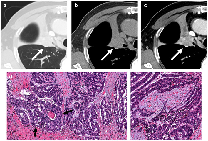
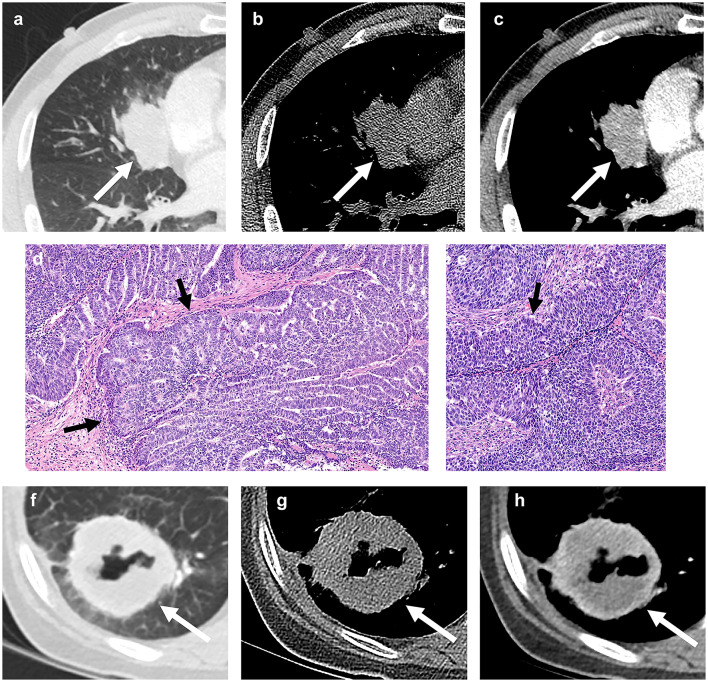
This study aimed to compare computed tomography (CT) findings between basaloid lung squamous cell carcinoma (SCC) and non-basaloid SCC. From July 2003 to April 2021, 39 patients with surgically proven basaloid SCC were identified. For comparison, 161 patients with surgically proven non-basaloid SCC from June 2018 to January 2019 were selected consecutively. Clinical features, demographic characteristics, and CT findings were compared using chi-square test or Fisher's exact test except for differences in means for which Student's t-test was used. Additionally, Mantel-Haenszel test was performed to control the confounding of the presence of cavitation between basaloid and non-basaloid SCCs with tumors stratified by clinical T staging. Compared with patients with non-basaloid SCC, patients with basaloid SCC had significantly (p < 0.001) more frequent respiratory symptoms at the time of presentation. Regarding CT findings, endobronchial tumor growth and obstructive pneumonia or atelectasis were significantly (p = 0.028 and p = 0.028, respectively) more common in basaloid SCC than in non-basaloid SCC. Compared with non-basaloid SCC, cavitation was absent (p = 0.005) and internal profuse necrosis was significantly (p = 0.022) less frequent in basaloid SCC. Furthermore, presence of cavitation consistently showed significant difference after the tumors stratified based on clinical T staging (p = 0.015). Basaloid SCC had some CT findings different from non-basaloid SCC. Basaloid SCC showed more frequent endobronchial tumor growth with obstructive pneumonia or atelectasis. Internal profuse necrosis was less common, and cavitation was absent in basaloid SCC compared to non-basaloid SCC.
Keywords: Computed tomography; Lung neoplasms; Squamous cell carcinoma.
© 2024. The Author(s).
Conflict of interest statement
Declarations. Competing interests: The authors declare no competing interests.
Figures
Similar articles
-
CT findings of basaloid squamous cell carcinoma of the lung in 12 patients: A distinct category of squamous cell carcinoma in 2015 WHO classification of lung tumors.Medicine (Baltimore). 2022 May 13;101(19):e29197. doi: 10.1097/MD.0000000000029197. Medicine (Baltimore). 2022. PMID: 35583530 Free PMC article.
-
CT radiomics analysis of lung cancers: Differentiation of squamous cell carcinoma from adenocarcinoma, a correlative study with FDG uptake.Eur J Radiol. 2020 Jul;128:109032. doi: 10.1016/j.ejrad.2020.109032. Epub 2020 Apr 26. Eur J Radiol. 2020. PMID: 32361604
-
High-resolution CT findings of primary lung cancer with cavitation: a comparison between adenocarcinoma and squamous cell carcinoma.Clin Radiol. 2016 Nov;71(11):1126-31. doi: 10.1016/j.crad.2016.06.110. Epub 2016 Jul 6. Clin Radiol. 2016. PMID: 27394062
-
Pulmonary squamous cell carcinoma and lymphoepithelial carcinoma - morphology, molecular characteristics and differential diagnosis.Histopathology. 2024 Jan;84(1):32-49. doi: 10.1111/his.15076. Epub 2023 Nov 7. Histopathology. 2024. PMID: 37936498 Review.
-
Esophageal Carcinosarcoma with Basaloid Squamous Cell Carcinoma: A Case Report and Review of the Literature.Tohoku J Exp Med. 2019 Dec;249(4):255-263. doi: 10.1620/tjem.249.255. Tohoku J Exp Med. 2019. PMID: 31852851 Review.
References
-
- Brambilla, E. et al. Basal cell (basaloid) carcinoma of the lung: a new morphologic and phenotypic entity with separate prognostic significance. Hum. Pathol.23, 993–1003. 10.1016/0046-8177(92)90260-a (1992). - PubMed
-
- Beasley, M. B., Brambilla, E. & Travis, W. D. The 2004 World Health Organization classification of lung tumors. Semin Roentgenol.40, 90–97. 10.1053/j.ro.2005.01.001 (2005). - PubMed
-
- Moro-Sibilot, D. et al. Lung carcinomas with a basaloid pattern: a study of 90 cases focusing on their poor prognosis. Eur. Respir J.31, 854–859. 10.1183/09031936.00058507 (2008). - PubMed
-
- Sturm, N. et al. Thyroid transcription factor 1 and cytokeratins 1, 5, 10, 14 (34betaE12) expression in basaloid and large-cell neuroendocrine carcinomas of the lung. Hum. Pathol.32, 918–925. 10.1053/hupa.2001.27110 (2001). - PubMed
-
- Brambilla, C. et al. Lung squamous cell carcinomas with basaloid histology represent a specific molecular entity. Clin. Cancer Res.20, 5777–5786. 10.1158/1078-0432.CCR-14-0459 (2014). - PubMed
Publication types
MeSH terms
LinkOut - more resources
Full Text Sources
Medical
Research Materials
Miscellaneous
