

Resolvin D1 combined with exercise rehabilitation alleviates neurological injury in mice with intracranial hemorrhage via the BDNF/TrkB/PI3K/AKT pathway
- PMID: 39733073
- PMCID: PMC11682414
- DOI: 10.1038/s41598-024-83019-w
Resolvin D1 combined with exercise rehabilitation alleviates neurological injury in mice with intracranial hemorrhage via the BDNF/TrkB/PI3K/AKT pathway
Abstract
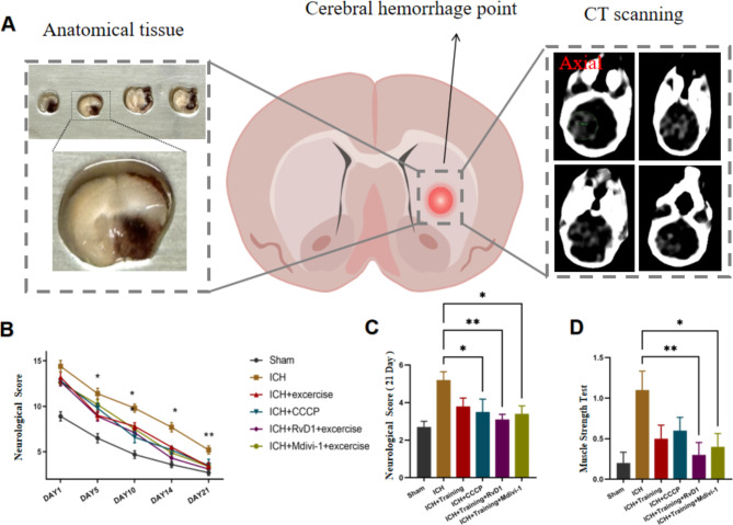
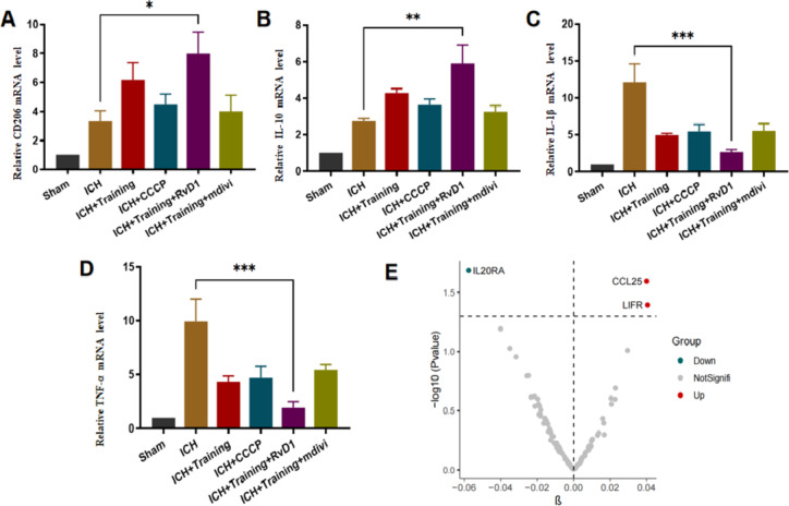
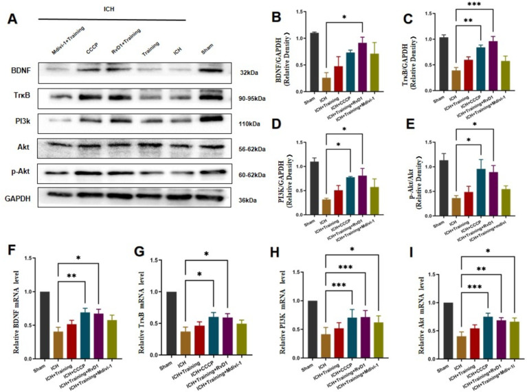
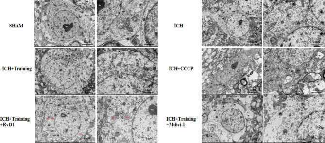
Resolvin D1 (RvD1) is an endogenous anti-inflammatory mediator that modulates the inflammatory response and promotes inflammation resolution. RvD1 has demonstrated neuroprotective effects in various central nervous system contexts; however, its role in the pathophysiological processes of intracerebral hemorrhage (ICH) and the potential protective mechanisms when combined with exercise rehabilitation remain unclear. A mouse model of ICH was established using collagenase, and treatment with RvD1 combined with three weeks of exercise rehabilitation significantly improved neurological deficits, muscle strength, learning, and memory in ICH mice while reducing anxiety-like behavior. RvD1 combined with exercise rehabilitation upregulated anti-inflammatory factors, inhibited the inflammatory state, and activated the BDNF/TrkB/PI3K/AKT pathway. TUNEL staining confirmed a decrease in residual apoptotic neurons, while transmission electron microscopy showed an increase in mitochondrial autophagosomes with combined treatment. Mendelian randomization and molecular docking further confirmed the association of RvD1 with targets related to mitophagy and inflammatory factors, clarifying the mechanism of RvD1 involvement. In summary, RvD1 combined with exercise rehabilitation activates the BDNF/TrkB/PI3K/AKT signaling pathway, effectively reduces neuronal apoptosis and inflammatory responses following ICH in mice, and participates in mitochondrial autophagy-related states. This comprehensive therapeutic strategy promotes neurological recovery and provides insights for clinical management of this condition.
Keywords: Inflammation; Intracerebral hemorrhage; Mitophagy; Neuronal Apoptosis; RvD1; exercise rehabilitation.
© 2024. The Author(s).
Conflict of interest statement
Declarations. ARRIVE quidelines statement: The study was carried out in compliance with the ARRIVE guidelines. Competing interests: The authors declare no competing interests.
Figures


References
Publication types
MeSH terms
Substances
Grants and funding
- 2024Y883/Scientific Research Foundation of Education Department of Yunnan Province
- 2024Y883/Scientific Research Foundation of Education Department of Yunnan Province
- 2024Y883/Scientific Research Foundation of Education Department of Yunnan Province
- 2024Y883/Scientific Research Foundation of Education Department of Yunnan Province
- 2024Y883/Scientific Research Foundation of Education Department of Yunnan Province
- 2024Y883/Scientific Research Foundation of Education Department of Yunnan Province
- 2024Y883/Scientific Research Foundation of Education Department of Yunnan Province
- FZ2023ZD028/Projects of the Research and Development Fund of Dali University
- FZ2023ZD028/Projects of the Research and Development Fund of Dali University
- FZ2023ZD028/Projects of the Research and Development Fund of Dali University
- FZ2023ZD028/Projects of the Research and Development Fund of Dali University
- FZ2023ZD028/Projects of the Research and Development Fund of Dali University
- 202101AN070028/Key project of the special fund for basic research of local undergraduate colleges and universities granted by Department of Science and Technology of Yunnan Province
- 202101AN070028/Key project of the special fund for basic research of local undergraduate colleges and universities granted by Department of Science and Technology of Yunnan Province
- 202101AN070028/Key project of the special fund for basic research of local undergraduate colleges and universities granted by Department of Science and Technology of Yunnan Province
- 202101AN070028/Key project of the special fund for basic research of local undergraduate colleges and universities granted by Department of Science and Technology of Yunnan Province
- 2019ZD036/Key Laboratory of medical molecular diagnosis in Colleges and Universities of Yunnan Province
- 2019ZD036/Key Laboratory of medical molecular diagnosis in Colleges and Universities of Yunnan Province
- 2019ZD036/Key Laboratory of medical molecular diagnosis in Colleges and Universities of Yunnan Province
LinkOut - more resources
Full Text Sources
