. 2024 Dec 28;14(1):31500.
doi: 10.1038/s41598-024-82959-7.

Histological effects of combined therapy involving scar resection, decellularized scaffolds, and human iPSC-NS/PCs transplantation in chronic complete spinal cord injury

Affiliations

Histological effects of combined therapy involving scar resection, decellularized scaffolds, and human iPSC-NS/PCs transplantation in chronic complete spinal cord injury

Keitaro Ito et al. Sci Rep. .

Abstract

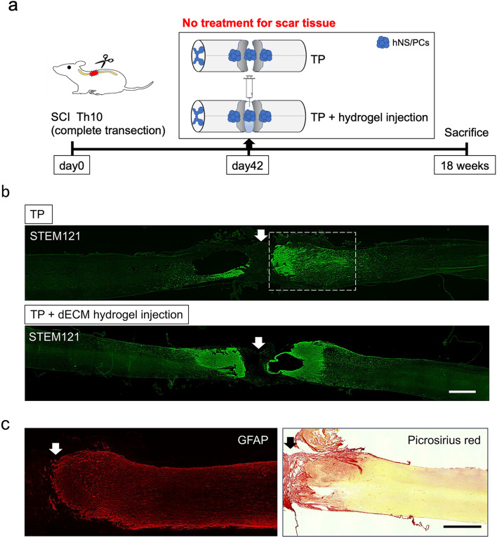
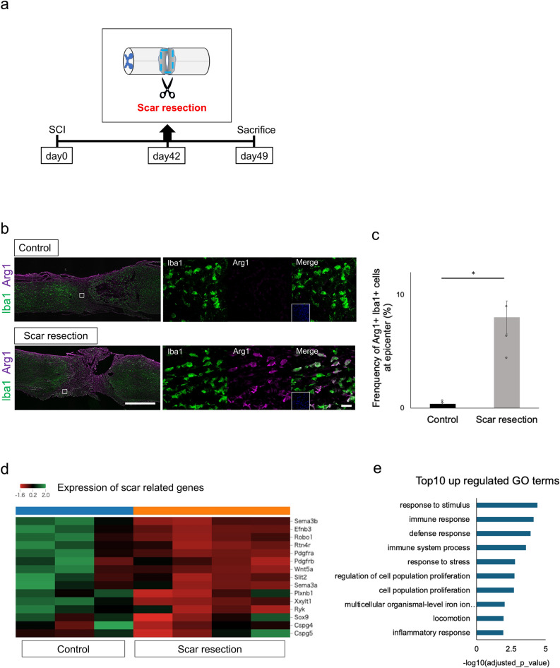
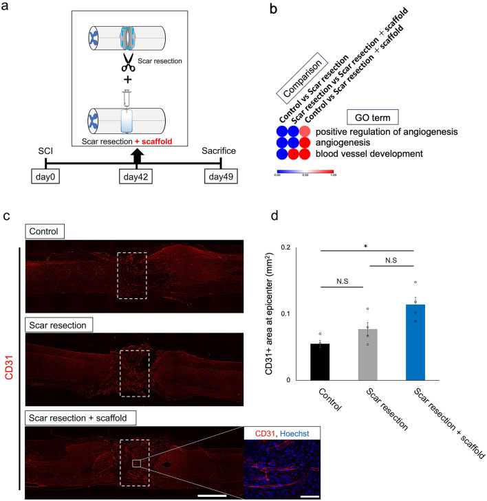
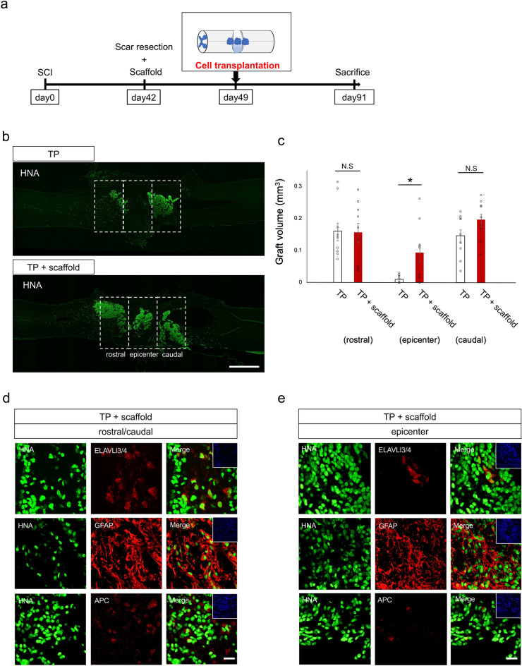
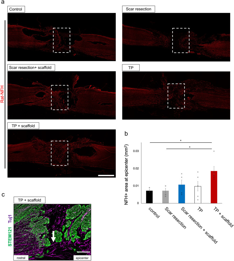
Chronic complete spinal cord injury (SCI) is difficult to treat because of scar formation and cavitary lesions. While human iPS cell-derived neural stem/progenitor cell (hNS/PC) therapy shows promise, its efficacy is limited without the structural support needed to address cavitary lesions. Our study investigated a combined approach involving surgical scar resection, decellularized extracellular matrix (dECM) hydrogel as a scaffold, and hNS/PC transplantation. To mitigate risks such as prion disease associated with spinal cord-derived dECM, we used kidney-derived dECM hydrogel. This material was chosen for its biocompatibility and angiogenic potential. In vitro studies with dorsal root ganglia (DRG) confirmed its ability to support axonal growth. In a chronic SCI rat model, scar resection enhanced the local microenvironment by increasing neuroprotective microglia and macrophages, while reducing inhibitory factors that prevent axonal regeneration. The combination of scar resection and dECM hydrogel further promoted vascular endothelial cell migration. These changes improved the survival of transplanted hNS/PCs and facilitated host axon regeneration. Overall, the integrated approach of scar resection, dECM hydrogel scaffolding, and hNS/PC transplantation has been proven to be a more effective treatment strategy for chronic SCI. However, despite histological improvements, no functional recovery occurred and further research is needed to enhance functional outcomes.

Keywords: Cell transplantation; Chronic phase; Scaffold; Scar resection; Spinal cord injury.


Conflict of interest statement

Declarations. Competing interests: The authors declare no competing interests.

Figures

Fig. 1
Fig. 1
Preparation and characterization of dECM hydrogels and effect of dECM hydrogel substrates on DRG neurite growth. (a) Diagram of the preparation process for kidney-derived dECM hydrogel. (b) SEM images of decellularized kidney hydrogel (8 mg/ml). Scale bar: 5 μm. (c) Experimental design and images of DRG culture on dECM hydrogel. (d) Tuj1-positive DRG neurites visualized using fluorescently stained images on different hydrogels. Scale bar: 200 μm. (e) Quantification of the neurite length of DRG on collagen I (n = 3) and dECM hydrogel (8 mg/ml (n = 4) and 16 mg/ml (n = 3)). One-way ANOVA followed by the Tukey–Kramer test; N.S., not significant; *P < 0.05. Data are expressed as mean ± SEM.
Fig. 2
Fig. 2
Therapeutic effects of dECM hydrogel injection and iPSC-NSPCs for chronic phase complete transected spinal cord. (a) Experimental design, images of the complete transection model, dECM hydrogel injection, and iPSC-NSPC transplantation in the chronic phase. TP, cell transplantation. (b) Representative STEM121-stained sagittal spinal cord sections at 12 weeks post-transplantation, showing non-viable transplanted cells in the epicenter. The arrows indicate the epicenter. Scale bar, 1000 μm. (c) Immunohistochemical analysis of the magnified boxed area from (b), illustrating the transplanted cells adhering to the cavity, glial scar (GFAP), and fibrous scar (picrosirius red stain). The arrows indicate the epicenter. Scale bar, 1000 μm.
Fig. 3
Fig. 3
Therapeutic effects of scar resection: immunohistochemical staining and RNA-seq analysis 7 days post-scar resection in the chronic transected SCI model. (a) Experimental design and images of scar resection in the chronic transected SCI model. (b) Representative midsagittal sections stained for Iba1 and Arg1 in the control and scar resection groups (enlarged images on the right). Scale bars, 1000 μm (left) and 20μm (right). (c) Percentages of the neuroprotective microglia and macrophages (Iba1 and Arg1) in the midsagittal sections (n = 4 each). Mann–Whitney U test; N.S., not significant; *P < 0.05. Data are expressed as mean ± SEM. (d) Heatmap of the axon inhibitor-associated and scar formation-related genes. Scale, z-score. (e) Enriched GO biological process terms increased in the scar resection group.
Fig. 4
Fig. 4
Therapeutic effects of scar resection and dECM hydrogel (scaffold). (a) Experimental design and images of scar resection and dECM hydrogel (scaffold) in the chronic transected SCI model. (b) Heatmap of the angiogenesis-associated genes in the scar resection and scar resection + scaffold groups. Scale, 0 <  − log 10|adjusted P| < 1.00. (c) Examination of neovascularization at the epicenter using immunohistochemical staining for CD31. Scale bar, 1000 μm. (d) Quantification of the CD31+ area in the 1000-μm-wide regions at the epicenter (n = 4 each). One-way ANOVA followed by the Tukey–Kramer test; N.S., not significant; *P < 0.05. Data are expressed as mean ± SEM.
Fig. 5
Fig. 5
Therapeutic effects of iPSC-NSPC transplantation after scar resection and scaffold. (a) Experimental design and images of iPSC-NSPC transplantation after scar resection and scaffold in the chronic phase. (b) Representative immunohistochemical HNA staining of the midsagittal sections. Scale bar, 1000 μm. (c) Quantification of the grafted cell volume in the transplantation and transplantation + scaffold groups (n = 9 each). Mann–Whitney U test; N.S., not significant; *P < 0.05. Data are expressed as mean ± SEM. (d, e) Representative images of the neural cells differentiated from the graft cells (HNA+), showing co-staining with ELAVL3/4 (neurons), GFAP (astrocytes), and APC (oligodendrocytes). Scale bars, 20 μm.
Fig. 6
Fig. 6
Combined therapy promoted the regeneration of neuronal fibers. (a) Examination of the host neurons around the lesion epicenter via immunohistochemical staining for rodent-specific NF-H. Scale bar, 1000 μm. (b) Quantification of the NF-H + area: control group (n = 4), scar resection + PBS group (n = 4), scar resection + scaffold group (n = 4), TP group (n = 5), and TP + scaffold group (n = 5). One-way ANOVA followed by the Tukey–Kramer test; N.S., not significant; *P < 0.05. Data are expressed as mean ± SEM. (c) Immunohistochemical analysis of the magnified arrow area from (b). Representative staining for Tuj and STEM121 in the midsagittal sections. The arrows indicate Tuj1-positive fiber. Scale bar, 200 μm.

Similar articles

References

    1. Nagoshi, N. et al. Phase I/II study of intrathecal administration of recombinant human hepatocyte growth factor in patients with acute spinal cord injury: A double-blind, randomized clinical trial of safety and efficacy. J. Neurotrauma37, 1752–1758 (2020). - PubMed
    1. Honmou, O. et al. Intravenous infusion of auto serum-expanded autologous mesenchymal stem cells in spinal cord injury patients: 13 case series. Clin. Neurol. Neurosurg.203, 106565 (2021). - PubMed
    1. Fehlings, M. G. et al. Rho inhibitor VX-210 in acute traumatic subaxial cervical spinal cord injury: Design of the SPinal Cord Injury Rho INhibition InvestiGation (SPRING) Clinical Trial. J. Neurotrauma35, 1049–1056 (2018). - PMC - PubMed
    1. Grossman, R. G. et al. A prospective, multicenter, phase I matched-comparison group trial of safety, pharmacokinetics, and preliminary efficacy of riluzole in patients with traumatic spinal cord injury. J. Neurotrauma31, 239–255 (2014). - PMC - PubMed
    1. Sugai, K. et al. First-in-human clinical trial of transplantation of iPSC-derived NS/PCs in subacute complete spinal cord injury: Study protocol. Regen. Ther.18, 321–333 (2021). - PMC - PubMed

Publication types

Substances