. 2024 Dec 30;12(1):275.
doi: 10.1186/s40168-024-01995-7.

Live Akkermansia muciniphila boosts dendritic cell retinoic acid synthesis to modulate IL-22 activity and mitigate colitis in mice

Affiliations

Live Akkermansia muciniphila boosts dendritic cell retinoic acid synthesis to modulate IL-22 activity and mitigate colitis in mice

Hongbin Liu et al. Microbiome. .

Erratum in

Abstract

Background: The interplay between gut microbiota and immune responses is crucial in ulcerative colitis (UC). Though Akkermansia muciniphila (Akk) shows therapeutic potential, the mechanisms remain unclear. This study sought to investigate differences in therapeutic efficacy among different forms or strains of Akk and elucidate the underlying mechanisms.

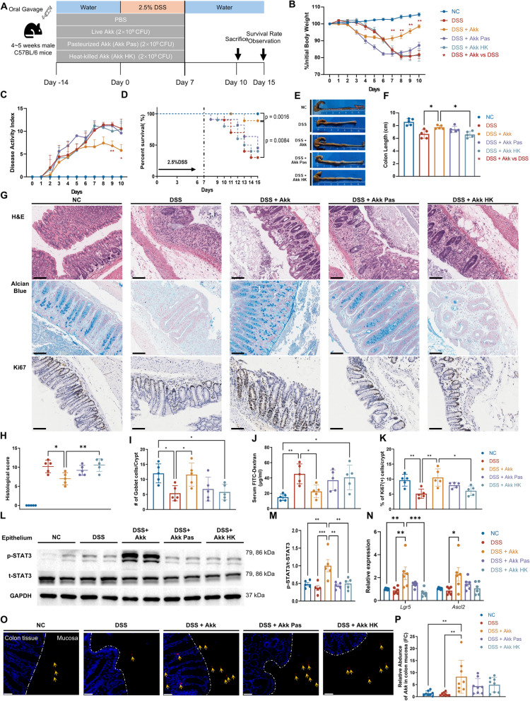
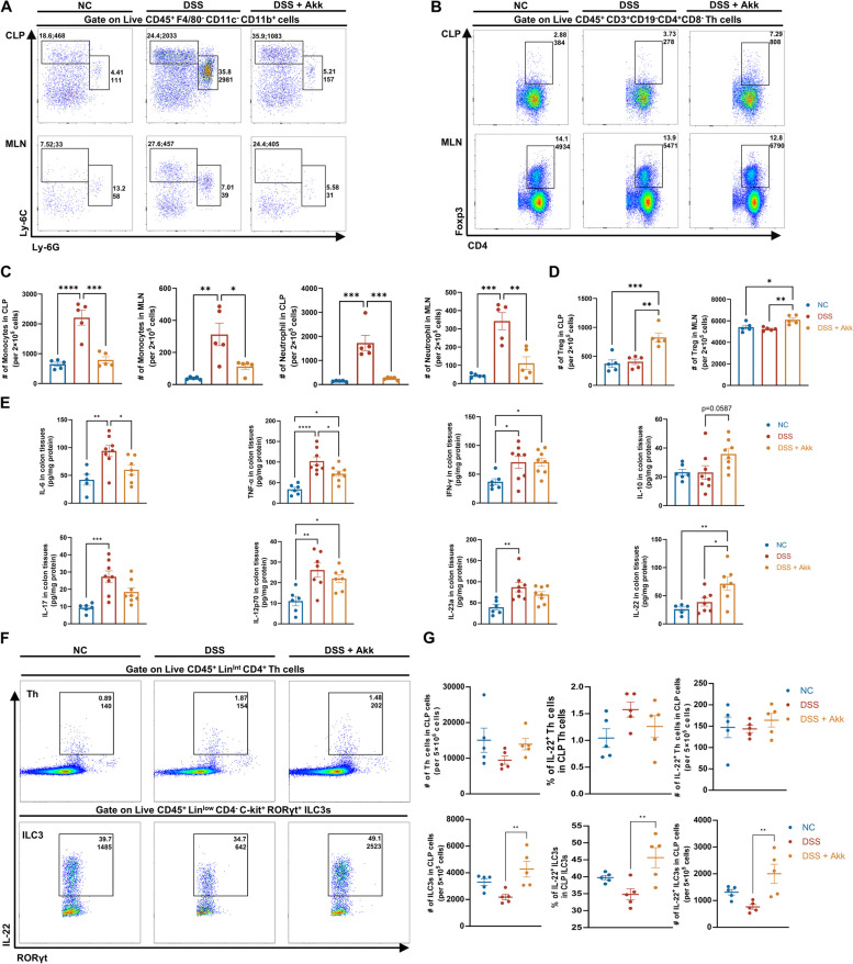
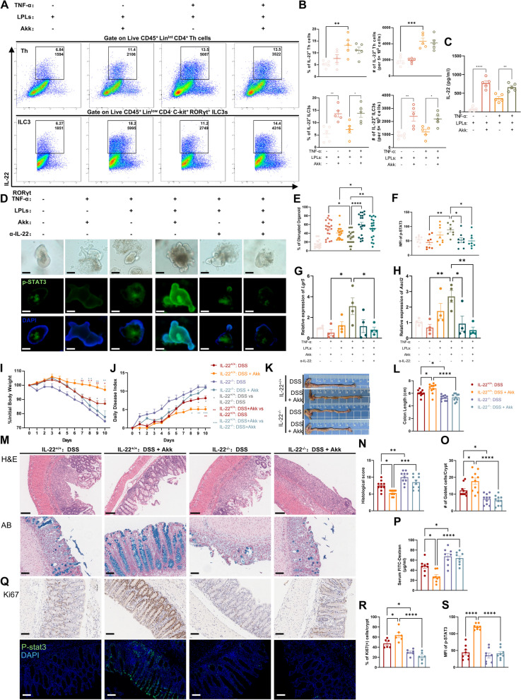
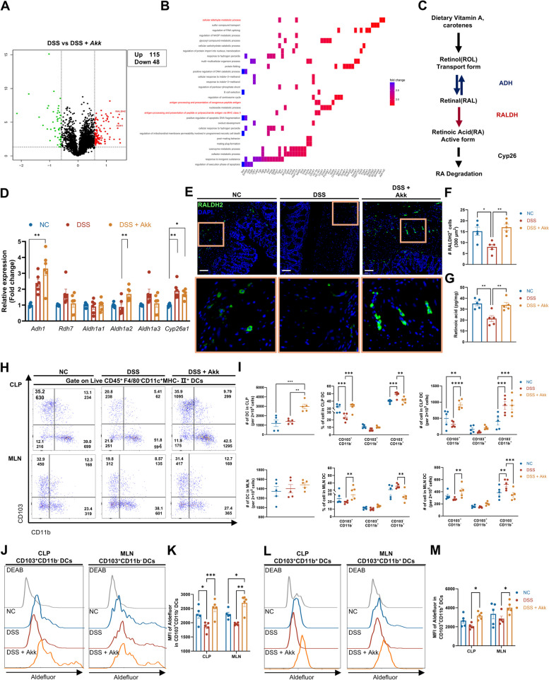
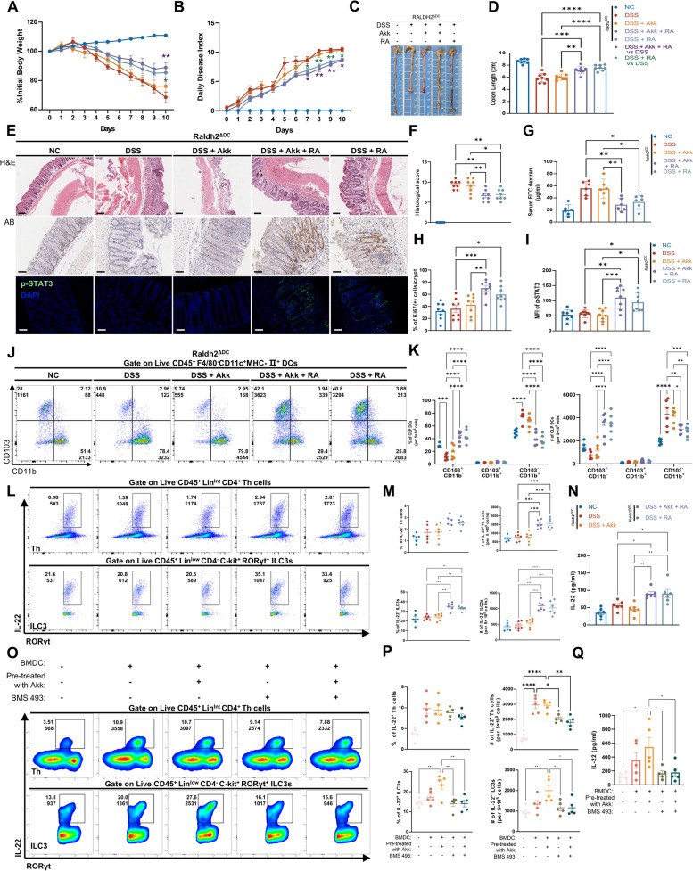
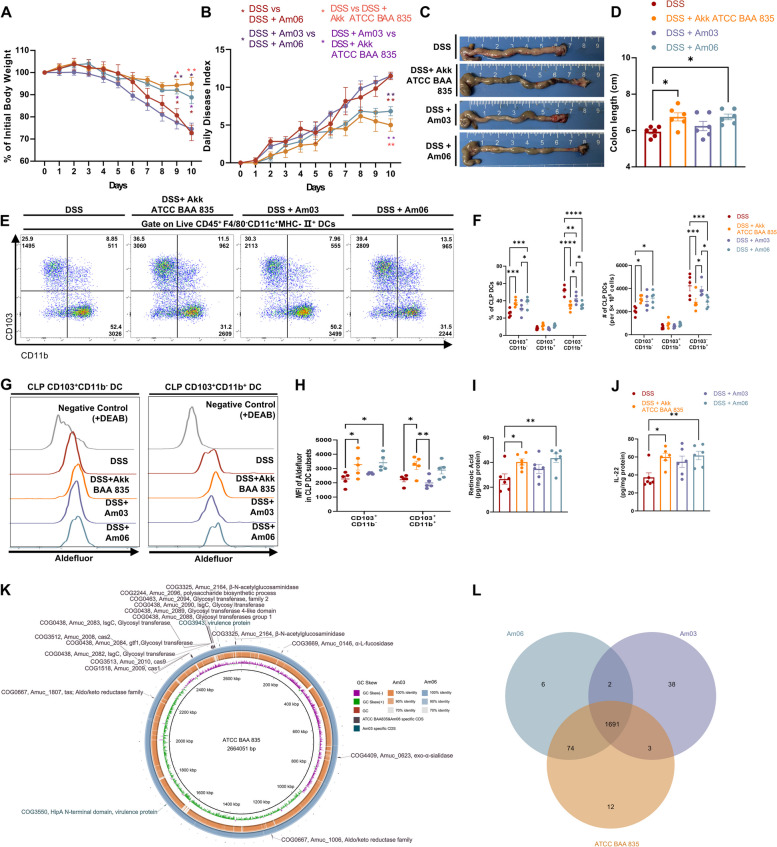
Results: Employing a dextran sulfate sodium (DSS)-induced colitis mouse model, we assessed Akk's impact on colitis using cellular cytokine analysis, immune phenotyping, proteomics, and biochemical methods. Our results suggest that treatment with live Akk effectively reduced colitis in the DSS-induced model, whereas heat-inactivated Akk did not yield the same results. Notably, Akk exhibited protective properties by promoting the secretion of IL-22 by Group 3 innate lymphoid cells (ILC3s), as evidenced by the absence of protection in IL-22 knockout mice. Additionally, Akk augmented the population of CD103+CD11b- dendritic cells (DCs) and enhanced their retinoic acid (RA) synthesis through the modulation of RALDH2, a crucial enzyme in RA metabolism. The depletion of RALDH2 in DCs diminished Akk's protective properties and impaired IL-22-mediated mucosal healing. Mechanistically, Akk activated RA production in DCs by enhancing the JAK2-STAT3 signaling pathway. Additionally, various strains of Akk may exhibit differing abilities to alleviate colitis, with the novel strain Am06 derived from breast milk showing consistent efficacy similar to the reference strain.

Conclusions: In summary, our findings indicate that certain strains of Akk may mitigate colitis through the promotion of RA synthesis and IL-22 secretion, underscoring the potential efficacy of Akk as a therapeutic intervention for the management of UC. Video Abstract.

Keywords: Akkermansia muciniphila; Dendritic cells; Group 3 innate lymphoid cells; IL-22; Retinoic acid; STAT3; Ulcerative colitis.


Conflict of interest statement

Declarations. Ethics approval and consent to participate: All animal experiments conducted adhered strictly to the Guidelines for Care and Use of Laboratory Animals as prescribed by the National Institutes of Health. Every animal study protocol utilized in this research was granted approval by the Institutional Animal Care and Use Committee at Southern Medical University (Approval No. K2019090). Consent for publication: Not applicable. Competing interests: The authors declare no competing interests.

Figures

Fig. 1
Fig. 1
Live Akk alleviates DSS-induced acute colitis. A Mice were assigned to receive either drinking water for 14 days followed by 2.5% DSS for 7 days and then drinking water again for 3 or 8 days for survival studies, or solely plain water for 24 or 29 days for survival analysis (n = 5; 10 for survival). Prior to and during DSS treatment, groups were orally administered live, pasteurized, or heat-killed Akk, or PBS. B–D Percentage of weight loss (B), disease activity index (DAI) (C), and survival status (D) were assessed daily for each treatment group. E, F Representative images of the colons (E) and the assessment of colon length (F) from different treatment groups. GI Representative images of the H&E-stained(upper), the Alcian Blue-stained colon sections (middle), and IHC images of Ki67 immunostaining (below) in the colon tissues of different treatment groups (scale bars 100 μm). Histological score (H) and goblet cells in the crypts (I) were assessed in the indicated groups. J Intestinal permeability of the relevant groups was determined by FITC-dextran level in serum. K Quantitative analysis of the percentage of Ki67+ cells in the crypts. L, M Representative immunoblot images of p-STAT3 and t-STAT3 in colon epithelial protein from different treatment groups (L). The protein levels of phosphorylated-STAT3 were quantified relative to total-STAT3 (M). N Expression of epithelial stem cell markers (Lgr5 and Ascl2) in the indicated groups (n = 8). O, P The relative abundance of Akk in the colonic mucosa was determined by q-PCR (O) (n = 8). Confocal microscopy of Akk (P): Bacteria (red) are highlighted by a yellow arrow; DAPI staining appears in blue (scale bar 50 μm). *P < 0.05, **P < 0.01, ***P < 0.001
Fig. 2
Fig. 2
Akk reduces colonic inflammation and upregulates IL-22 expression by ILC3 cells. AD Representative flow plots of CLP (top) and MLN (bottom) monocytes, neutrophils (A), and Tregs (B) in untreated mice and acute DSS-treated mice with gavage of PBS or Akk. Frequencies and absolute numbers of these specific populations (C&D) were determined (n = 5). E Cytokine levels in colonic proteins from relevant groups were assessed by ELISA (n = 6–8). F, G FACS analysis of total numbers of ILC3 cells (F), the frequencies and absolute numbers of CLP IL-22+ ILC3s and IL-22+ Th cells from the indicated mice (G) (n = 5). *P < 0.05, **P < 0.01, ***P < 0.001, ****P < 0.0001
Fig. 3
Fig. 3
The protective effect of Akk is mediated by the secretion of IL-22 by ILC3 cells. AC Colonic LPLs were treated with or without TNF-α or Akk (n = 5) for 12 h. The representative image of flow cytometric analysis for IL-22+ ILC3 cells and IL-22+ Th cells (A), including the frequencies and absolute numbers of these specific populations among different treatment groups (B). The IL-22 expression of cultured supernatants in these four groups was determined by ELISA (C). DH Organoids were cultured with or without Akk or LPLs in the presence or absence of TNF-α or anti-IL-22 for 12 h. The morphologies of organoids (upper) and the immunofluorescence analysis of p-STAT3 (below; p-STAT3 staining in green, DAPI staining in blue; scale bars 100 μm) were evaluated by light or fluorescence microscopy (D). The relative number of organoids with altered morphology (E) (n = 4, randomly select five fields of view for observation in each well) and the average fluorescence intensity of p-STAT3 (F) were quantitatively assessed (n = 8 organoids per group). The mRNA levels of Lgr5 (G) and Ascl2 (H) in colonic organoids from different treatment groups were determined (n = 4). IJ Littermate IL-22−/− or IL-22+/+ mice (n = 10) were assigned to receive either drinking water for 14 days followed by 2.5% DSS for 7 days and then drinking water again for 3 days. Prior to and during DSS treatment, groups were orally administered live Akk or PBS. The changes in body weight (I) and the disease activity index (J) were monitored daily following the DSS challenge. K, L Representative images of the colons from these mice (K), and measurement of colon length of the indicated mice (L). MO Representative images of H&E stained (upper, scale bars 200 μm) and Alcian blue (below) stained colon sections from relevant groups (scale bars 100 μm). Histological scores of colitis (N), goblet cells in the crypt (O), and intestinal permeability (P) were quantitatively assessed. QS Representative images of immunohistochemistry (IHC) staining for Ki67 (upper) and immunofluorescence images of p-STAT3 staining (below) (Q) in the colon tissues from different treatment groups (scale bars 100 μm); The quantitative analysis of the number of ki67+ cells in the crypt (R) and the average median fluorescent intensity (MFI) of p-STAT3 (S) were assessed using ImageJ. *P < 0.05, **P < 0.01, ***P < 0.001, ****P < 0.0001
Fig. 4
Fig. 4
Akk promotes RA synthesis in CD103+ DCs in mice with colitis. A, B Volcano plot (A) and KEGG enrichment pathway analysis (B) display differentially expressed proteins and associated pathways among acute DSS-treated mice with gavage of PBS or Akk (n = 5). C Schematic diagram illustrating retinoic acid metabolism. Vitamin A undergoes two-step oxidation reactions mediated by retinol dehydrogenase (ADH) and retinal dehydrogenase (RALDH) to produce retinoic acid, which can further undergo degradation by cytochrome enzymes such as CY26A1. D Expression of retinoic acid metabolism-related genes was evaluated in bulk colon samples from untreated mice and acute DSS-treated mice with gavage of PBS or Akk (n = 6). E, F Representative immunofluorescence images of RALDH2 immunostaining (E) in colon tissues from relevant groups (scale bars 100 μm). Orange boxes represent zoomed-in views. The average number of RALDH2+ cells per 300 μm2 (F) was determined from 5 randomly selected fields of view (n = 5). G ELISA test of whole colon homogenates to evaluate retinoic acid production (n = 5). H, I Representative images (H), frequency, and quantitative analysis (I) of total and different DC subsets in the CLP (upper) and MLN (below) by flow cytometry (n = 5). JM Representative images and quantitative analysis for FACS Aldefluor assay of CD103+CD11b DCs (J, K) and CD103+CD11b+ DCs (L, M) within the CLP and MLN (n = 5). *P < 0.05, **P < 0.01, ***P < 0.001
Fig. 5
Fig. 5
Retinoic acid synthesis in DCs is essential for Akk-induced IL-22 production in the inflamed colon. AD The pathology of colitis in littermate Raldh2ΔDC mice was assessed by observing untreated controls and test groups exposed to acute DSS with individual or combined treatments of Vehicle, Akk, or RA. Body weight changes (A), DAI (B), and colon lengths (C&D) were utilized to evaluate the pathology (n = 6). E, F Representative images of the H&E-stained(upper, scale bars 200 μm), the IHC images of Ki67 immunostaining colon sections (middle), and immunofluorescence images of p-STAT3 immunostaining (below) in the colon tissues of different treatment groups (scale bars 100 μm). Histological scores (F) were assessed in the indicated groups. G Intestinal permeability of the relevant groups was determined by FITC-dextran level in serum (n = 6). H Quantitative analysis of the percentage of ki67+ cells in the crypts by ImageJ (n = 6). I The average fluorescence intensity of p-STAT3 was quantitatively assessed from relevant groups (n = 6). J, K Representative images (J) and quantitative analysis (K) of different CLP DC subsets from various experimental groups were obtained through flow cytometry (n = 5). L, M Representative images (L) and quantitative analysis (M) of IL-22+ Th cells and IL-22+ ILC3s in the CLP from various experimental groups were conducted via flow cytometry (n = 5). N The IL-22 production of the relevant groups was assessed by conducting ELISA tests on whole colon homogenates (n = 6). OQ BMDCs, pre-treated with or without Akk, were then co-cultured with colonic LPLs, in the presence or absence of the RA pan-receptor inhibitor BMS493. Representative images (O), Representative flow plots (O), and both frequencies and quantitative analyses (P) of IL-22+ ILC3s and IL-22+ Th cells from relevant cells were conducted using flow cytometry. The IL-22 expression of co-cultured supernatants in relevant groups was determined by ELISA (Q) (n = 5). *P < 0.05, **P < 0.01, ***P < 0.001, ****P < 0.0001
Fig. 6
Fig. 6
Akk promotes RA synthesis in DCs via the JAK2-STAT3 signaling pathway. AF Representative images and quantitative analysis of Flow-cytometric analysis for p-STAT3 staining in the CLP total DCs (A, B), CD103+CD11b DCs (C, D), CD103+CD11b+ DCs (E, F) from untreated mice and acute DSS-treated mice with gavage of PBS or Akk (n = 5–6). G A correlation analysis was performed on total and CD103+ CLP DC subsets to determine the relationship between the MFI of p-STAT3 and Aldefluor (n = 17). H, I GM-CSF derived BMDCs were treated with LPS or Akk alone or together for 12 h. Representative immunoblot images of TLR2, p-JAK2, JAK2, p-STAT3, t-STAT3, SOCS2, p-mTOR, and mTOR in different treatment groups. Relative protein levels of indicated proteins were quantified to GAPDH. The protein levels of phosphorylated-STAT3 were quantified relative to total-STAT3 (n = 5). J, K GM-CSF derived BMDCs were treated with LPS or Akk alone or together, in the absence or presence of STAT3 inhibitor Stattic for 12 h. Representative immunoblot images of TLR2, p-STAT3, and t-STAT3 in different treatment groups. Relative protein levels for these proteins were calibrated to GAPDH while p-STAT3 levels were relative to total-STAT3 (n = 6). L, M Representative images (L) and quantitative analysis (M) of FACS Aldefluor assay for these indicated cells (n = 5). N The activity of the RALDH2 promoter was assessed by dual-luciferase reporter assay in BMDCs transfected with the vector or STAT3 overexpression (STAT3 OE) plasmids. O Schematic images of the potential STAT3 binding sites in the promoter of RALDH2 predicted by JASPAR (upper). ChIP analysis of STAT3 occupancy at the RALDH2 promoter in STAT3-overexpressing BMDCs (below). *P < 0.05, **P < 0.01, ***P < 0.001, ****P < 0.0001
Fig. 7
Fig. 7
Akk exhibits strain-specific effects in ameliorating acute colitis in mice. AD The pathology of colitis was evaluated among the 4 acute colitis groups with PBS, Akk ATCC BAA-835, Am03, or Am06 gavage respectively. Body weight changes (A), DAI (B), and colon lengths (C, D) were utilized to evaluate the pathology (n = 6). E, F Representative images (E), frequencies, and quantitative analysis (F) of different CLP DC subsets from relevant groups were assessed by Flow cytometry (n = 5). G, H Representative images (G) and quantitative analysis (H) for FACS Aldefluor assay of CLP CD103+CD11b DCs and CD103+CD11b+ DCs from relevant groups (n = 5). I, J ELISA test of whole colon homogenates to evaluate RA (I) and IL-22 (J) production from relevant groups (n = 6). K Genomic comparison of Akk ATCC BAA-835, Am03, or Am06 chromosomes, starting from the predicted replication origin, includes (1) Am03 or Am06 contigs, (2) ATCC BAA-835 genome identity based on BLASTN against Am03 or Am06, and (3) interested strain-specific coding sequences (CDSs) for Am03, along with unique CDSs for ATCC BAA-835 and Am06, with annotated functions. L Visualization of the Eggnog output comparing the number of unique and shared orthologs of Akk BAA 835, Am03, and Am06. *P < 0.05, **P < 0.01, ***P < 0.001, ****P < 0.0001

Similar articles

Cited by

References

    1. Le Berre C, Honap S, Peyrin-Biroulet L. Ulcerative colitis. The Lancet. 2023;402:571–84. - PubMed
    1. Glymenaki M, Singh G, Brass A, Warhurst G, McBain AJ, Else KJ, et al. Compositional changes in the gut mucus microbiota precede the onset of colitis-induced inflammation. Inflamm Bowel Dis. 2017;23:912–22. - PubMed
    1. van de Guchte M, Mondot S, Doré J. Dynamic properties of the intestinal ecosystem call for combination therapies, targeting inflammation and microbiota, in ulcerative colitis. Gastroenterology. 2021;161:1969-1981.e12. - PubMed
    1. Gilliland A, Chan JJ, De Wolfe TJ, Yang H, Vallance BA. Pathobionts in Inflammatory Bowel Disease: Origins, Underlying Mechanisms, and Implications for Clinical Care. Gastroenterology. 2024;166:44–58. - PubMed
    1. Jia DJC, Wang QW, Hu YY, He JM, Ge QW, Qi YD, et al. Lactobacillus johnsonii alleviates colitis by TLR1/2-STAT3 mediated CD206+ macrophagesIL-10 activation. Gut Microbes. 2022;14:2145843. - PMC - PubMed

Supplementary concepts

LinkOut - more resources